. 2010 Mar 18:4:8.
doi: 10.3389/fnbeh.2010.00008. eCollection 2010.

Behavioral and neurobiological changes in C57BL/6 mouse exposed to cuprizone: effects of antipsychotics

Affiliations

Behavioral and neurobiological changes in C57BL/6 mouse exposed to cuprizone: effects of antipsychotics

Haiyun Xu et al. Front Behav Neurosci. .

Abstract

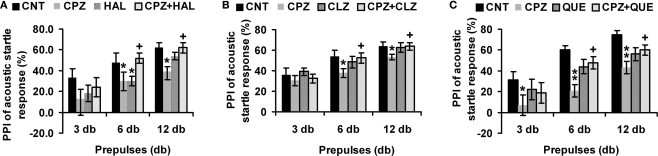
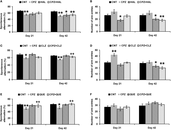
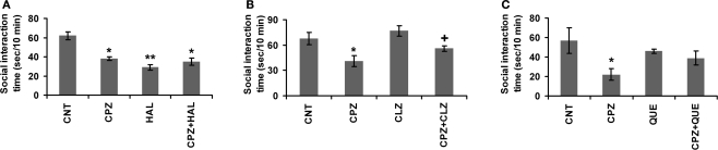
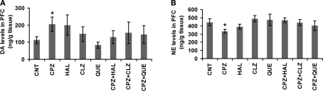
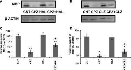
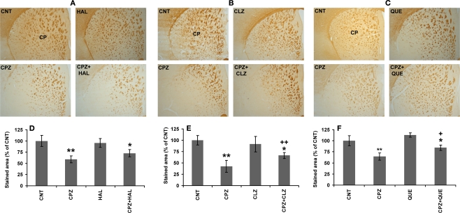
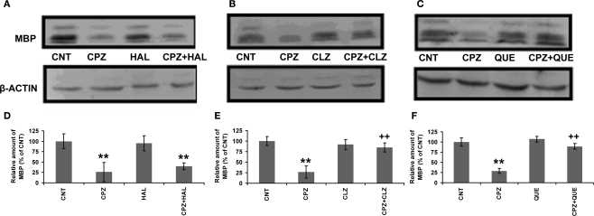
Recent human studies suggest a role for altered oligodendrocytes in the pathophysiology of schizophrenia. Our recent animal study has reported some schizophrenia-like behaviors in mice exposed to cuprizone (Xu et al., 2009), a copper chelator that has been shown to selectively damage the white matter. This study was to explore mechanisms underlying the behavioral changes in cuprizone-exposed mice and to examine effects of the antipsychotics haloperidol, clozapine and quetiapine on the changes in the mice. Mice given cuprizone for 14 days showed a deficit in the prepulse inhibition of acoustic startle response and higher dopamine in the prefrontal cortex (PFC), which changes were not seen in mice given cuprizone plus antipsychotics. Mice given cuprizone for 21 days showed lower spontaneous alternations in Y-maze, which was not seen in mice treated with cuprizone plus the antipsychotics. Mice given cuprizone for 28 days displayed less social interactions, which was not seen in mice given cuprizone plus clozapine/quetiapine, but was seen in mice given cuprizone plus haloperidol. Mice given cuprizone for 42 days showed myelin sheath loss and lower myelin basic protein in PFC, caudate putamen, and hippocampus. The white matter damage in PFC was attenuated in mice given cuprizone plus clozapine/haloperidol. But the white matter damage in caudate putamen and hippocampus was only attenuated by clozapine and quetiapine, not by haloperidol. These results help us to understand the behavioral changes and provide experimental evidence for the protective effects of antipsychotics on white matter damage in cuprizone-exposed mice.

Keywords: MBP; antipsychotics; behavior; mouse; oligodendrocytes; schizophrenia model.


Figures

Figure 1
Figure 1
Effects of antipsychotics on the CPZ-induced deficits in PPI test. Control and experimentally treated C57BL/6 mice were subjected to PPI test on the same day (14th day after CPZ-exposure). (A) The data of the HAL experiment. (B) The data of the CLZ experiment. (C) The data of the QUE experiment. Data were expressed as M ± SEM (n = 6 to 12/group). CNT, Control group; CPZ, Cuprizone group; CLZ, Clozapine group; HAL, Haloperidol group; QUE, Quetiapine group; CPZ + CLZ, mice received both cuprizone and clozapine; CPZ + HAL, mice received both cuprizone and haloperidol; CPZ + QUE, mice received both cuprizone and quetiapine. PPI = Prepulse inhibition; db = Decibel. *p < 0.05, **p < 0.01, compared to the CNT group; +p < 0.05, compared to the CPZ group.
Figure 2
Figure 2
Effects of antipsychotics on the CPZ-induced abnormal performance in the Y-maze test. Control and experimentally treated C57BL/6 mice were subjected to Y-maze test on the same days (21st and 42nd days after CPZ-exposure). (A) The data of the spontaneous alternation in the HAL experiment. (B) The data of the number of arm entries in the HAL experiment. (C) The data of the spontaneous alternation in the CLZ experiment. (D) The data of the number of arm entries in the CLZ experiment. (E) The data of the spontaneous alternation in the QUE experiment. (F) The data of the number of arm entries in the QUE experiment. Data were expressed as M ± SEM (n = 6 to 12/group). *p < 0.05, **p < 0.01, compared to the CNT group; ++p < 0.01, compared to the CPZ group.
Figure 3
Figure 3
Effects of antipsychotics on the CPZ-induced deficits in the social interaction. Control and experimentally treated C57BL/6 mice were subjected to social interaction test on the same day (28th day after CPZ-exposure). (A) The data of the HAL experiment. (B) The data of the CLZ experiment. (C) The data of the QUE experiment. Data were expressed as M ± SEM (n = 6 pairs/group). *p < 0.05, **p < 0.01, compared to the CNT group; +p < 0.05, compared to the CPZ group.
Figure 4
Figure 4
Effects of antipsychotics on the CPZ-induced changes in levels of DA and NE in PFC. (A) Control and experimentally treated C57BL/6 mice were sacrificed on 14th day after CPZ-exposure. DA levels in PFC were measured by means of HPLC. (B) Control and experimentally treated C57BL/6 mice were sacrificed on 21st day after CPZ-exposure. NE levels in PFC were measured by means of HPLC. Data were expressed as M ± SEM (ng/g tissue; n = 6 to 12/group). *p < 0.05, compared to the CNT group.
Figure 5
Figure 5
Effects of HAL and CLZ on the CPZ-induced decrease in MBP in PFC. Control and experimentally treated C57BL/6 mice were sacrificed on 43rd day after CPZ-exposure. The PFC was dissected out of the brain and processed for Western-blot analysis to measure MBP levels. The upper photographs (A) and (B) are representative Western-blots from HAL and CLZ experiments, respectively. The bar charts (C) and (D) in the bottom panel are the statistical results of the amount of MBP relative to β-actin in the same corresponding lanes as labeled. Data were expressed as M ± SEM (n = 6/group). *p < 0.05, **p < 0.01, compared to the CNT group. +p < 0.05, compared to the CPZ group.
Figure 6
Figure 6
CLZ and QUE, but not HAL, protect the CPZ-induced myelin sheath loss in CP. (A) Images showing MBP-like immunoreactivity (MBP-LI) in CP of mice in the HAL experiment. (B) Images showing MBP-LI in CP of mice in the CLZ experiment. (C) Images showing MBP-LI immunoreactivity in CP of mice in the QUE experiment. (D) The bar chart of MBP-LI area in tissue sections of CP in the HAL experiment. (E) The bar chart of MBP-LI area in tissue sections of CP in the CLZ experiment. (F) The bar chart of MBP-LI area in tissue sections of CP in the QUE experiment. Data were expressed as M ± SEM (n = 6/group). *p < 0.05, **p < 0.01, compared to the CNT group. +p < 0.05, ++p < 0.01, compared to the CPZ group.
Figure 7
Figure 7
Effects of antipsychotics on the CPZ-induced decrease in MBP in CP. Control and experimentally treated C57BL/6 mice were sacrificed on 43rd day after CPZ-exposure. The CP was dissected out of the brain and processed for Western-blot analysis to measure MBP levels. The upper photographs (A–C) are representative Western-blots from the HAL, CLZ, and QUE experiments, respectively. The bar charts (D–F) in the bottom panel are the statistical results of the amount of MBP relative to β-actin in the same corresponding lanes as labeled. Data were expressed as M ± SEM (n = 6/group). **p < 0.01, compared to the CNT group; ++p < 0.01, compared to the CPZ group.

Similar articles

Cited by

References

    1. Abi-Dargham A., Rodenhiser J., Printz D., Zea-Ponce Y., Gil R., Kegeles L. S., Weiss R., Cooper T. B., Mann J. J., Van Heertum R. L., Gorman J. M., Laruelle M. (2000). Increased baseline occupancy of D2 receptors by dopamine in schizophrenia. Proc. Natl. Acad. Sci. U.S.A. 97, 8104–810910.1073/pnas.97.14.8104 - DOI - PMC - PubMed
    1. Ardekani B. A., Nierenberg J., Hoptman M. J., Javitt D. C., Lim K. O. (2003). MRI study of white matter diffusion anisotropy in schizophrenia. Neuroreport 14, 2025–202910.1097/00001756-200311140-00004 - DOI - PubMed
    1. Bai O., Chlan-Fourney J., Bowen R., Keegan D., Li X. M. (2003). Expression of brain-derived neurotrophic factor mRNA in rat hippocampus after treatment with antipsychotic drugs. J. Neurosci. Res. 71, 127–13110.1002/jnr.10440 - DOI - PubMed
    1. Bai O., Zhang H., Li X. M. (2004). Antipsychotic drugs clozapine and olanzapine upregulate bcl-2 mRNA and protein in rat frontal cortex and hippocampus. Brain Res. 1010, 81–8610.1016/j.brainres.2004.02.064 - DOI - PubMed
    1. Bassett A. S., Chow E. W., AbdelMalik P., Gheorghiu M., Husted J., Weksberg R. (2003). The schizophrenia phenotype in 22q11 deletion syndrome. Am. J. Psychiatry 160, 1580–158610.1176/appi.ajp.160.9.1580 - DOI - PMC - PubMed