

The Drosophila neural lineages: a model system to study brain development and circuitry
- PMID: 20306203
- PMCID: PMC2886914
- DOI: 10.1007/s00427-010-0323-7
The Drosophila neural lineages: a model system to study brain development and circuitry
Abstract
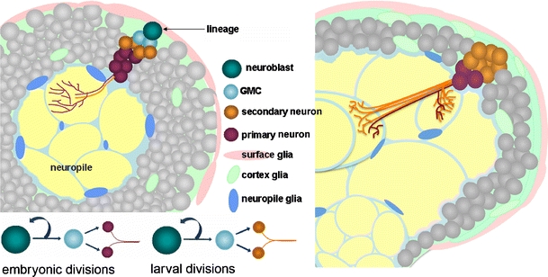
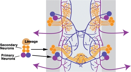
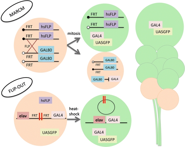
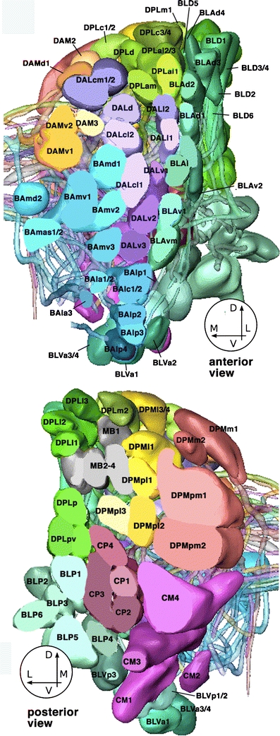
In Drosophila, neurons of the central nervous system are grouped into units called lineages. Each lineage contains cells derived from a single neuroblast. Due to its clonal nature, the Drosophila brain is a valuable model system to study neuron development and circuit formation. To better understand the mechanisms underlying brain development, genetic manipulation tools can be utilized within lineages to visualize, knock down, or over-express proteins. Here, we will introduce the formation and development of lineages, discuss how one can utilize this model system, offer a comprehensive list of known lineages and their respective markers, and then briefly review studies that have utilized Drosophila neural lineages with a look at how this model system can benefit future endeavors.
Figures


Similar articles
-
Postembryonic development of transit amplifying neuroblast lineages in the Drosophila brain.Neural Dev. 2009 Dec 11;4:44. doi: 10.1186/1749-8104-4-44. Neural Dev. 2009. PMID: 20003348 Free PMC article.
-
Neural lineages of the Drosophila brain: a three-dimensional digital atlas of the pattern of lineage location and projection at the late larval stage.J Neurosci. 2006 May 17;26(20):5534-53. doi: 10.1523/JNEUROSCI.4708-05.2006. J Neurosci. 2006. PMID: 16707805 Free PMC article.
-
Identification of Drosophila type II neuroblast lineages containing transit amplifying ganglion mother cells.Dev Neurobiol. 2008 Aug;68(9):1185-95. doi: 10.1002/dneu.20648. Dev Neurobiol. 2008. PMID: 18548484 Free PMC article.
-
Wiring the Drosophila Brain with Individually Tailored Neural Lineages.Curr Biol. 2017 Jan 23;27(2):R77-R82. doi: 10.1016/j.cub.2016.12.026. Curr Biol. 2017. PMID: 28118595 Review.
-
Neuroblast formation and patterning during early brain development in Drosophila.Bioessays. 2004 Jul;26(7):739-51. doi: 10.1002/bies.20062. Bioessays. 2004. PMID: 15221856 Review.
Cited by
-
Functional analysis of conserved sequences within a temporally restricted neural precursor cell enhancer.Mech Dev. 2011 Mar-Apr;128(3-4):165-77. doi: 10.1016/j.mod.2011.02.001. Epub 2011 Feb 16. Mech Dev. 2011. PMID: 21315151 Free PMC article.
-
Asymmetric activity of NetrinB controls laterality of the Drosophila brain.Nat Commun. 2023 Feb 24;14(1):1052. doi: 10.1038/s41467-023-36644-4. Nat Commun. 2023. PMID: 36828820 Free PMC article.
-
The labial gene is required to terminate proliferation of identified neuroblasts in postembryonic development of the Drosophila brain.Biol Open. 2012 Oct 15;1(10):1006-15. doi: 10.1242/bio.20121966. Epub 2012 Aug 17. Biol Open. 2012. PMID: 23213378 Free PMC article.
-
Simple model systems reveal conserved mechanisms of Alzheimer's disease and related tauopathies.Mol Neurodegener. 2023 Nov 10;18(1):82. doi: 10.1186/s13024-023-00664-x. Mol Neurodegener. 2023. PMID: 37950311 Free PMC article. Review.
-
Nutritional regulation of stem and progenitor cells in Drosophila.Development. 2013 Dec;140(23):4647-56. doi: 10.1242/dev.079087. Development. 2013. PMID: 24255094 Free PMC article. Review.
References
-
- Bate CM. Embryogenesis of an insect nervous system. I. A map of the thoracic and abdominal neuroblasts in Locusta migratoria. J Embryol Exp Morphol. 1976;35:107–123. - PubMed
Publication types
MeSH terms
Grants and funding
LinkOut - more resources
Full Text Sources
Other Literature Sources
Molecular Biology Databases
