Review
. 2010 Jul;67(13):2251-69.
doi: 10.1007/s00018-010-0327-5. Epub 2010 Mar 21.

The perpetual movements of anaphase

Affiliations
Review

The perpetual movements of anaphase

Helder Maiato et al. Cell Mol Life Sci. 2010 Jul.

Abstract

One of the most extraordinary events in the lifetime of a cell is the coordinated separation of sister chromatids during cell division. This is truly the essence of the entire mitotic process and the reason for the most profound morphological changes in cytoskeleton and nuclear organization that a cell may ever experience. It all occurs within a very short time window known as "anaphase", as if the cell had spent the rest of its existence getting ready for this moment in an ultimate act of survival. And there is a good reason for this: no space for mistakes. Problems in the distribution of chromosomes during cell division have been correlated with aneuploidy, a common feature observed in cancers and several birth defects, and the main cause of spontaneous abortion in humans. In this paper, we critically review the mechanisms of anaphase chromosome motion that resisted the scrutiny of more than 100 years of research, as part of a tribute to the pioneering work of Miguel Mota.


Figures

Fig. 1
Fig. 1
a First illustration of anaphase during the division of the first micromeres in the worm Rhynchelmis by A. Kowalevski (adapted from [2]). b Contemporary view of micromere formation in the sand dollar embryo (courtesy of George von Dassow, University of Washington, WA, USA). c Newt lung cell in anaphase as viewed by fluorescence microscopy (courtesy of Conly Rieder, Wadsworth Center, NY, USA). d Sequence of the anaphase movement in a plant cell as originally depicted by Strasburger (adapted from [4]). e Sequence of the anaphase movement from a time-lapse movie of a Drosophila S2 cell stably expressing mCherry-α-tubulin (red) and GFP-H2B-Histone (green) (courtesy of Sara Moutinho-Pereira, IBMC, University of Porto, Portugal). Note the simultaneity of anaphase A and B
Fig. 2
Fig. 2
Schematic representation of anaphase models. a Traction-fiber model where active microtubule depolymerization of k-fibers by kinesin-13 proteins occurs at the minus ends. b Kinetochore dynein model where dynein ATPase activity drives anaphase A; dynein at the poles and cortex provides a pulling force that prevents spindle collapse and may drive anaphase B. c Conformational wave model where the bending of protofilaments at microtubule plus ends coupled with kinetochore fibrils drives anaphase A independently of ATP hydrolysis. d Kinetochore pac-man model where active microtubule plus end depolymerization by kinesin-13 proteins occurs at kinetochores. e Spindle matrix model where immobilized kinesin motors exert a poleward force on k-fibers. f Actin/myosin model (blue lines) where actin is a structural component of k-fibers which contracts by the action of myosin. g Coupled sliding model where motors of the kinesin family slide interpolar microtubules leading to spindle elongation and whose poleward force is transmitted along k-fibers by microtubule cross-linking molecules. Combinations between different models are possible
Fig. 3
Fig. 3
Hans Ris (right) chairs the session where Miguel Mota (left) presented his view of the kinetochore as an “engine” in the International Genetics Symposia held in Japan in 1956. Mota used to fly his own Piper Cub from Lisbon to Porto to use the Electron Microscope. His passion for aeroplanes evidently had a strong influence in the conceptualization of his anaphase hypothesis
Fig. 4
Fig. 4
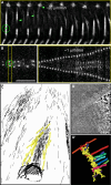
a Laser-mediated severing of a k-fiber in a Drosophila S2 cell stably expressing GFP-α-tubulin during metaphase. Note the fast depolymerization of the pole-proximal fragment and that the chromosome which remains attached to the severed k-fiber maintains its equatorial position (adapted from [52]). b Laser-mediated severing of the centromeric region in a Drosophila S2 cell stably expressing CID-GFP during metaphase. Note the slow poleward migration of each daughter kinetochore after surgery. Scale bar (a,b) 5 μm. c 3D-electron microscope reconstruction of a severed k-fiber from crane flies after irradiation with a UV microbeam (adapted from [177]). Non-kinetochore microtubules in the vicinity of the resulting k-fiber stub were pseudocolored in yellow. d Single slice from a tomographic reconstruction of a Ptk1 kinetochore showing both end-on and lateral MT binding (courtesy from Yimin Dong and Bruce McEwen, Wadsworth Center, NY, USA). Scale bar 100 nm. d’ 3D surface rendering of the 3D volume of the same kinetochore
Fig. 5
Fig. 5
a, b Time-lapse sequences of LLC-PK cells stably expressing GFP-α-tubulin after treatment with DMSO or 20 μM Cytochalasin D, respectively. Chromosomes can be visualized by differential interference contrast in the superimposed image. Time between frames = 1 min. c, d Distributions of measurements of k-fiber shortening and half-spindle elongation velocities after treatment with DMSO or 20 μM Cytochalasin D. The mean k-fiber shortening velocities in DMSO and Cytochalasin D were, respectively, 0.19 ± 0.13 (n = 6 cells) and 0.26 ± 0.17 (n = 4 cells) for the represented period. Error intervals represent standard deviation. The differences are not statistically significant (P = 0.296; t test). The median half-spindle elongation velocities in the same cells after DMSO or Cytochalasin D were, respectively, 0.47 ± 0.12 and 0.60 ± 0.21. Error intervals represent standard deviation. The differences are not statistically significant (P = 0.058; Mann–Whitney rank sum test)

References

    1. Baker J. The cell-theory: a restatement, history, and critique: Part V. The multiplication of nuclei. Q J Microscop Sci. 1955;3–96:449–481.
    1. Kowalevski A. Embryologische Studien an Wurmerm und Arthropoden. Mem Acad Imper Sci St. Petersburg. 1871;16:1–70.
    1. Flemming W. Beiträge zur Kenntniss der Zelle und ihrer Lebenserscheinungen. Arch Mikr Anat. 1879;16:302–436. doi: 10.1007/BF02956386. - DOI
    1. Strasburger E. Die Controversen der indirecten Kerntheilung. Arch Mikr Anat. 1884;23:246–304. doi: 10.1007/BF02952312. - DOI
    1. Carter LA. The somatic mitosis of Stegomyia fasciata . Q J Microscop Sci. 1918;2–63:375–386.

Publication types

LinkOut - more resources