Review
. 2011 Jun;41(1):7-27.
doi: 10.1007/s00726-010-0552-0. Epub 2010 Mar 22.

Cysteine S-conjugate β-lyases: important roles in the metabolism of naturally occurring sulfur and selenium-containing compounds, xenobiotics and anticancer agents

Affiliations
Review

Cysteine S-conjugate β-lyases: important roles in the metabolism of naturally occurring sulfur and selenium-containing compounds, xenobiotics and anticancer agents

Arthur J L Cooper et al. Amino Acids. 2011 Jun.

Abstract

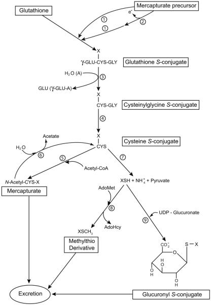
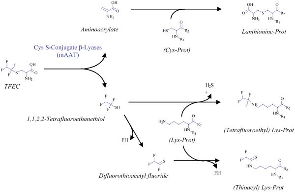
Cysteine S-conjugate β-lyases are pyridoxal 5'-phosphate-containing enzymes that catalyze β-elimination reactions with cysteine S-conjugates that possess a good leaving group in the β-position. The end products are aminoacrylate and a sulfur-containing fragment. The aminoacrylate tautomerizes and hydrolyzes to pyruvate and ammonia. The mammalian cysteine S-conjugate β-lyases thus far identified are enzymes involved in amino acid metabolism that catalyze β-lyase reactions as non-physiological side reactions. Most are aminotransferases. In some cases the lyase is inactivated by reaction products. The cysteine S-conjugate β-lyases are of much interest to toxicologists because they play an important key role in the bioactivation (toxication) of halogenated alkenes, some of which are produced on an industrial scale and are environmental contaminants. The cysteine S-conjugate β-lyases have been reviewed in this journal previously (Cooper and Pinto in Amino Acids 30:1-15, 2006). Here, we focus on more recent findings regarding: (1) the identification of enzymes associated with high-M(r) cysteine S-conjugate β-lyases in the cytosolic and mitochondrial fractions of rat liver and kidney; (2) the mechanism of syncatalytic inactivation of rat liver mitochondrial aspartate aminotransferase by the nephrotoxic β-lyase substrate S-(1,1,2,2-tetrafluoroethyl)-L-cysteine (the cysteine S-conjugate of tetrafluoroethylene); (3) toxicant channeling of reactive fragments from the active site of mitochondrial aspartate aminotransferase to susceptible proteins in the mitochondria; (4) the involvement of cysteine S-conjugate β-lyases in the metabolism/bioactivation of drugs and natural products; and (5) the role of cysteine S-conjugate β-lyases in the metabolism of selenocysteine Se-conjugates. This review emphasizes the fact that the cysteine S-conjugate β-lyases are biologically more important than hitherto appreciated.


Figures

Fig. 1
Fig. 1
The mercapturate pathway and associated side reactions. If the mercapturate precursor contains an electrophilic center it may react directly with GSH (reaction 1). Alternatively, the precursor may be converted to a compound with an electrophilic center (reaction 2) prior to reaction with GSH. Reactions 1 through 5 represent the mercapturate pathway. Reactions 7, 8 and 9 are alternative reactions for elimination of the cysteine S-conjugate. Reactions 7 plus 8 denote the thiomethyl shunt. The thiomethyl compound (XSCH3) may be excreted unchanged or further oxidized to sulfoxide, sulfone or CO2 and sulfate, which are excreted. For some cysteine S-conjugates metabolism may also involve conversion to the α-keto acid, α-hydroxy acid, oxidatively decarboxylated product or sulfoxide (not shown). Enzymes: 1) glutathione S-transferases, 2) oxidases that generate an electrophilic center for attack by GSH (in some cases oxidation may be non-enzymatic); 3) γ-glutamyltransferase (GGT); 4) dipeptidases; 6) aminoacylases; 7) cysteine S-conjugate β-lyases; 8) thiomethyltransferase; 9) UDP-glucuronosyltransferases. In vivo the hydrolysis reaction of GGT predominates over the formation of γ-glutamyl amino acids. Abbreviations: AdoHcy, S-adenosyl-L-homocysteine; AdoMet, S-adenosyl-L-methionine; A, amino acid, dipeptide or GSH acceptor for the γ-glutamyltransferase reaction; γ-GLU-A, γ-glutamyl amino acid (or γ-glutamyldipeptide; γ-glutamylglutathione). Many potentially toxic xenobiotics and a few endogenous compounds are metabolized through the mercapturate pathway. Modified in part from Silbernagl and Heuner (1993) and Cooper and Pinto (2008). Reproduced from Cooper and Hanigan (2010) with permission.
Fig 2
Fig 2
Syncatalytic inactivation of mAAT by TFEC. A. Inactivation of mAAT (0.1 mg/ml) in the presence of 10 mM sodium phosphate buffer (pH 7.5), 10 mM α-ketoglutarate and 10 mM TFEC; 10°C. Enzyme activities (●) are expressed as percent relative to the activity of a sample of native enzyme maintained under identical conditions, but in the absence of TFEC. B. Mass spectra of mAAT. Mass spectra were obtained before (blue line) and after 4 h of reaction with TFEC (red line). The protein aliquots were desalted in a C8 reverse phase column and injected into the mass spectrometer. Mass spectra were deconvoluted using the algorithm included in the Xcalibur software. For the covalently modified protein the masses are given as increment in mass relative to the mass of the mAAT polypeptide (44,587 daltons).
Fig. 3
Fig. 3
LC/ESI mass spectral analysis of covalently modified peptides. Tandem mass spectra of the triply-charged peptide-ions at m/z of 1006.4 (122FFK~FSRDVFLPK~ PSWGNHTIPF148R), 603.31 (242HFIEQGINVC@LCQSYAK^NM*GLYGE266R) and 981.30 (267VGAFTVVC@ KDAEEAK282R). Modified amino acid residues are indicated as in Table 4.
Fig. 4
Fig. 4
Reaction pathways of TFEC with mAAT. Aminoacrylate reacts with sulfhydryl groups of cysteine (Cys) residues resulting in the formation of lanthionine residues. The other product, tetrafluoroethanethiol, can react directly or after decomposition to difluorothioacetyl fluoride, with the ε-amino group of K residues. In the first case, loss of H2S results in the formation of a tetrafluoroethyl derivative. In the second case, loss of FH results in the formation of a thioacyl derivative.
Fig. 5
Fig. 5
Three dimensional structure of mAAT indicating residues modified by covalent attachment of TFEC fragments. The relative positions of the modified residues are shown in green on the native structure of mAAT. The PLP coenzyme is shown in yellow forming a Schiff base with the Lys 258. The diagram was generated using the program VMD (Theoretical Biophysics Group, University of Illinois at Urbana-Champaign) and the X-ray structures of mAAT (pyridoxal form, Protein Data Bank entry code 1TAT) from chicken heart mitochondria.
Fig. 6
Fig. 6
A. Mitochondrial toxicant channeling in vivo. TFEC (R) transport into the mitochondria results in conversion to the thioacylating agent (Rδ+) by β-lyases [e.g. mAAT (1), shown here as a homodimer]. Co-immunoprecipitation and biochemical studies confirm the close juxtaposition of other DFTAL target proteins – especially those of energy metabolism [KGDHC (2) and mitACON (3)]. Considerable evidence suggests that BCATm can form a metabolon with the branched-chain α-keto acid dehydrogenase complex (BCDHC) (Islam et al., 2010 and references quoted therein). Since BCATm can catalyze a β-lyase reaction with TFEC (Cooper et al., 2003) it is probable that subunits of the BCDHC are also inactivated by toxicant channeling. Other complexes not known to be associated with any aminotransferase/cysteine S-conjugate β-lyase activities [e.g. PDC] are not modified or inactivated by thioacylation. The curved arrow represents “self-thioacylation” of mAAT (syncatalytic inactivation). Positions of outer (OM) and inner (IM) mitochondrial membranes are indicated. Adapted from Cooper et al. (2002a). B. TFEC-induced mitochondrial pathophysiology. Submitochondrial fractionation studies confirm a unified movement of matrix, IM and inter-membrane space DFTAL-labeled and unmodified proteins to the periphery of rat renal mitochondria isolated from kidney tissue treated with TFEC. Such movements are consistent with a permeability transition and Δψi collapse as assessed by morphological changes, flow cytometry and biochemical inhibitor studies (see the text for details). Note that DFTAL covalent modifications of mAAT, KGDHC components, mitACON, HSP70, and HSP60 were detected in kidney mitochondria of rats exposed in vivo to TFEC by immunochemical methods. This finding does not, however, preclude the possibility of additional covalent modifications occurring in vivo as has been demonstrated for mAAT in vitro as shown in Fig. 4. Adapted from Bruschi et al. (1993).
Fig. 7
Fig. 7
De novo synthesis of stress responsive protein cytosolic HSP70i following TFEC exposure (100, 200 or 300 μM; 4h) to TAMH cells in cell culture. A. Quantitative real-time PCR determination of hsp70i mRNA with gene-specific primers. Data are represented as the fluorescence yield from successive PCR reaction cycles with the fold-induction indicated to the right (maximum of ≈ 400×). Data are the mean (± s.d.) from 3 independent experiments (n = 3-6 replicate plates per experiment). B. New synthesis of HSP70i protein also in TAMH cell cultures in vitro which paralleled hsp70i mRNA induction. Migration of Mr standards are shown to the left. Original and some additional (unpublished) data are from the study of Ho et al. (2006).
Fig. 8
Fig. 8
Proposed mechanism for the metabolic conversion of busulfan to tetrahydrothiophene (THT) and to a glutathione adduct containing a lanthionine bridge. Facile elimination of a methanesulfonyl (II) group from busulfan (I) occurs by nucleophilic attack of cysteine (Cys) or glutathione (GSH), resulting in the formation of thioether conjugates [cysteine S-conjugate (III) and glutathione S-conjugate (IV), respectively]. Conversion of I to IV may occur non-enzymatically or may be accelerated by various GSTs. Structures III and IV spontaneously undergo elimination/ring closure to generate sulfonium conjugates [cysteine S-conjugate (V) and glutathione S-conjugate (VI), respectively]. Structure VI is converted to V by the action of γ-glutamyltransferase (a) and dipeptidases (b), both of which possess broad specificities. V undergoes a facile β-elimination reaction to yield THT (VII), ammonium (IX) and pyruvate (X), which may be catalyzed by OH-, PLP or several PLP-containing enzymes. THT (VII) may also be generated from γ-E-THT-AG (VI) by a non-enzymatic β-elimination reaction that results in the formation of γ-glutamyldehydroalanylglycine (EDAG) (VIII). Note that the sulfur in THT is not derived from busulfan, but rather from the sulfur of GSH. VIII contains an activated vinyl group and spontaneously undergoes a Michael reaction with GSH to form structure XI that contains a lanthionine bridge. Modified from Cooper et al. (2008b) and Younis et al. (2008)

Similar articles

Cited by

References

    1. Abraham DG, Cooper AJL. Glutamine transaminase K and cysteine S-conjugate β-lyase activity stains. Anal Biochem. 1991;197:421–427. - PubMed
    1. Abraham DG, Cooper AJL. Cloning and expression of a rat kidney cytosolic glutamine transaminase K that has strong sequence homology to kynurenine-pyruvate aminotransferase. Arch Biochem Biophys. 1996;335:311–320. - PubMed
    1. Abraham DG, Patel PP, Cooper AJL. Isolation from rat kidney of a high molecular weight cysteine S-conjugate β-lyase with activity toward leukotriene E4. J Biol Chem. 1995a;270:180–188. - PubMed
    1. Abraham DG, Thomas RJ, Cooper AJL. Glutamine transaminase K is not a major cysteine S-conjugate β-lyase of rat kidney mitochondria: Evidence that a high-molecular-weight enzyme fulfills this role. Mol Pharmacol. 1995b;48:855–860. - PubMed
    1. Adams B, Lowpetch K, Thorndycroft F, Whyte SM, Young DW. Stereochemistry of reactions of the inhibitor/substrates L- and D-chloroalanine with β-mercaptoethanol catalysed by L-aspartate aminotransferase and D-amino acid aminotransferase respectively. Org Biomol Med. 2005;3:3357–3364. - PubMed

Publication types