. 2010 Apr 27;4(4):2109-23.
doi: 10.1021/nn1000222.

Plasmonic nanobubbles as transient vapor nanobubbles generated around plasmonic nanoparticles

Affiliations

Plasmonic nanobubbles as transient vapor nanobubbles generated around plasmonic nanoparticles

Ekaterina Lukianova-Hleb et al. ACS Nano. .

Abstract

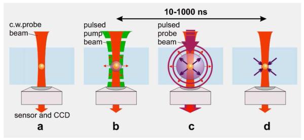
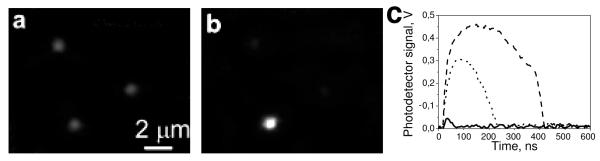
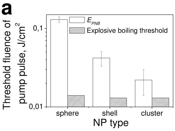
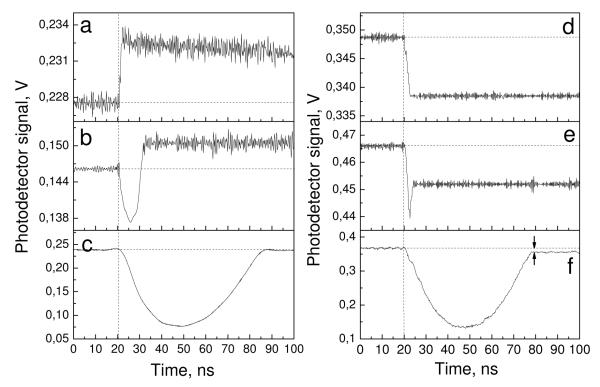
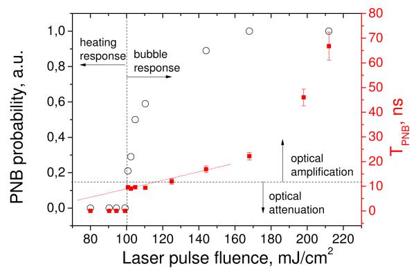
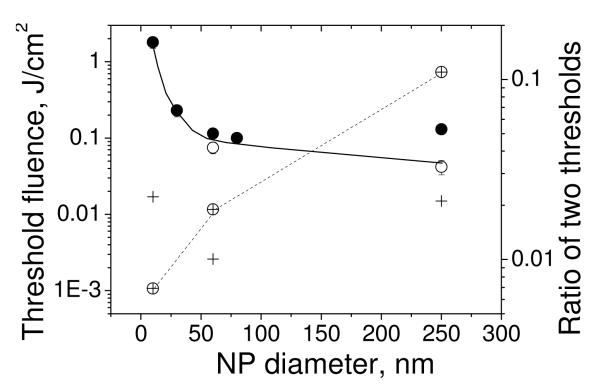
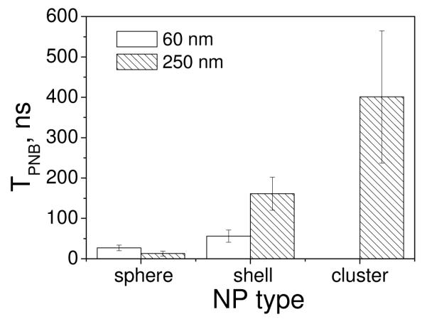
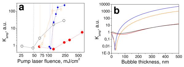
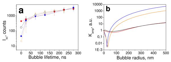
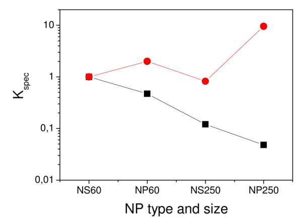
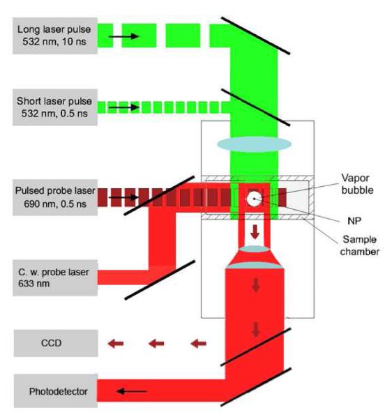
We have used short laser pulses to generate transient vapor nanobubbles around plasmonic nanoparticles. The photothermal, mechanical, and optical properties of such bubbles were found to be different from those of plasmonic nanoparticle and vapor bubbles, as well. This phenomenon was considered as a new complex nanosystem-plasmonic nanobubble (PNB). Mechanical and optical scattering properties of PNB depended upon the nanoparticle surface and heat capacity, clusterization state, and the optical pulse length. The generation of the PNB required much higher laser pulse fluence thresholds than the explosive boiling level and was characterized by the relatively high lower threshold of the minimal size (lifetime) of PNB. Optical scattering by PNB and its diameter (measured as the lifetime) has been varied with the fluence of laser pulse, and this has demonstrated the tunable nature of PNB.


Figures

Figure 1
Figure 1
Optical generation and detection of plasmonic nanobubbles: (a) gold NP in water is imaged with a low power probe laser beam (633 nm); (b) NP is exposed to a short pump laser pulse at specific fluence above (0.5 ns, 532 nm), that at the first stage produces the vapor layer around heated NP; (c) at the second stage when the pressure inside the vapor layer exceeds the outer pressure the vapor begins to expand into a bubble, which is detected with the two probe lasers (red and purple): scattering by the bubble forms the image and the time response; (d) a bubble collapses at the end of its lifespan (10-1000 ns).
Figure 2
Figure 2
Optical pulsed side-scattering images of (a) three 250 nm gold spheres in water, (b) transient vapor bubble generated around one NP positioned to the center of pump laser beam, (c) optical scattering time responses at the level of pump pulse fluence (0.13 J/cm2) for 250 nm gold sphere (solid), 250/15 nm silica-gold shell (dot) and for the cluster of gold shells (dash).
Figure 3
Figure 3
Observed thresholds for the bubble generation (EPNB) and estimated thresholds for explosive boiling around single gold NPs (250 nm solid sphere and silica-gold shell) and around shell clusters. Data are shown for a single excitation pump laser pulse (0.5 ns, 532 nm).
Figure 4
Figure 4
Time responses for single 80-nm gold nanoparticles (a-c) and molecular solution of Trypan Blue (d-f) obtained with a single pump laser pulse (vertical dashed line) near the PNB threshold fluences. Two arrows in f show the deviation of the after bubble signal from the baseline due to residual heating of the surrounding water.
Figure 5
Figure 5
Dependence of the PNB lifetime (TPNB, red) and the generation probability (black) around single 90 nm gold spheres upon the fluence of a single pump laser pulse (0.5 ns, 532 nm); vertical line separates the types of corresponding time responses (Figure 4) and the horizontal line separates the optical scattering effect.
Figure 6
Figure 6
Threshold fluences of pump laser pulse (0.5 ns, 532 nm) for the explosive boiling (+ − calculated for spheres ) and bubble generation (measured for ● – spheres and ○ – shells,) as functions of the diameter of gold NPs, solid line shows the bubble generation threshold trend, dashed line shows the trend for the ratio of explosive boiling threshold to bubble generation threshold (right Y axis).
Figure 7
Figure 7
Lifetimes of the PNBs (TPNB) generated around individual NPs (gold spheres and shells of 60 nm and 250 nm diameter) and around NP (shell) clusters at the fluence of the pump laser pulse 0.13 J/cm2.
Figure 8
Figure 8
Experimental (a) and theoretical (b) data on optical scattering by the nanoparticle-bubble system: (a) optical scattering amplification coefficient as function of pump pulse (0.5 ns, 532 nm) fluence: hollow black circle – 250 nm shells, red circle – 250 nm spheres, blue circle – 60 nm spheres, hollow orange circle – 60 nm shells, dashed vertical lines show the corresponding bubble generation thresholds; (b) optical scattering amplification coefficient calculated as the function of the bubble thickness (the difference in radii of the PNB and NP).
Figure 9
Figure 9
Experimental (a) and theoretical (b) data on optical scattering by the nanoparticle-bubble system: (a) pixel image amplitude of the bubble measured as function of the bubble lifetime (TPNB): optical scattering amplification coefficient as function of pump pulse (0.5 ns, 532 nm) fluence: hollow black circle – 250 nm shells, red circle – 250 nm spheres, blue circle – 60 nm spheres, hollow orange circle – 60 nm shells; (b) calculated optical scattering cross section of the nanoparticle-bubble system as function of the bubble radius.
Figure 10
Figure 10
Optical contrast coefficients for 60 nm shell (black squares)) and the corresponding PNB (red circles) generated around it at 0.13 J/cm2 as compared to other NPs (NS60 - 60 nm shells, NP60 – 60 nm spheres, NS250 - 250 nm shells, NP250 - 250 nm spheres) and the PNBs generated around them under identical conditions.
Figure 11
Figure 11
Experimental setup: single gold NPs in water were placed in the sample chamber mounted on the microscope stage; bubble generation was provided by focused single pulses (532 nm, 05 ns or 10 ns); a pulsed probing laser (690 nm, 05 ns) provided time-resolved optical scattering imaging of bubble and a continuous probing laser (633 nm, 1 mW) provided the monitoring of the integral optical scattering of bubble.

References

    1. Liao H, Nehl C, Hafner J. Biomedical Applications of Plasmon Resonant Metal Nanoparticles. Nanomedicine. 2006;1:201–208. - PubMed
    1. Pitsillides M, Joe E, Wei X, Anderson R, Lin C. Selective Cell Targeting with Light-Absorbing Microparticles and Nanoparticles. Biophys. J. 2003;84:4023–4032. - PMC - PubMed
    1. Loo C, Lowery A, Halas N, West J, Drezek R. Immunotargeted Nanoshells for Integrated Cancer Imaging and Therapy. Nano Lett. 2005;5:709–711. - PubMed
    1. Tong L, Zhao Y, Huff T, Hansen M, Wei A, Cheng J. Gold Nanorods Mediate Tumor Cell Death by Compromising Membrane Integrity. Adv. Mater. 2007;19:3136–3141. - PMC - PubMed
    1. El-Sayed I, Huang X, El-Sayed M. Selective Laser Photo-Thermal Therapy of Epithelial Carcinoma Using Anti-EGFR Antibody Conjugated Gold Nanoparticles. Cancer Lett. 2006;239:129–135. - PubMed

Publication types