Review
. 2010:28:491-533.
doi: 10.1146/annurev.immunol.021908.132550.

Molecular basis of calcium signaling in lymphocytes: STIM and ORAI

Affiliations
Review

Molecular basis of calcium signaling in lymphocytes: STIM and ORAI

Patrick G Hogan et al. Annu Rev Immunol. 2010.

Abstract

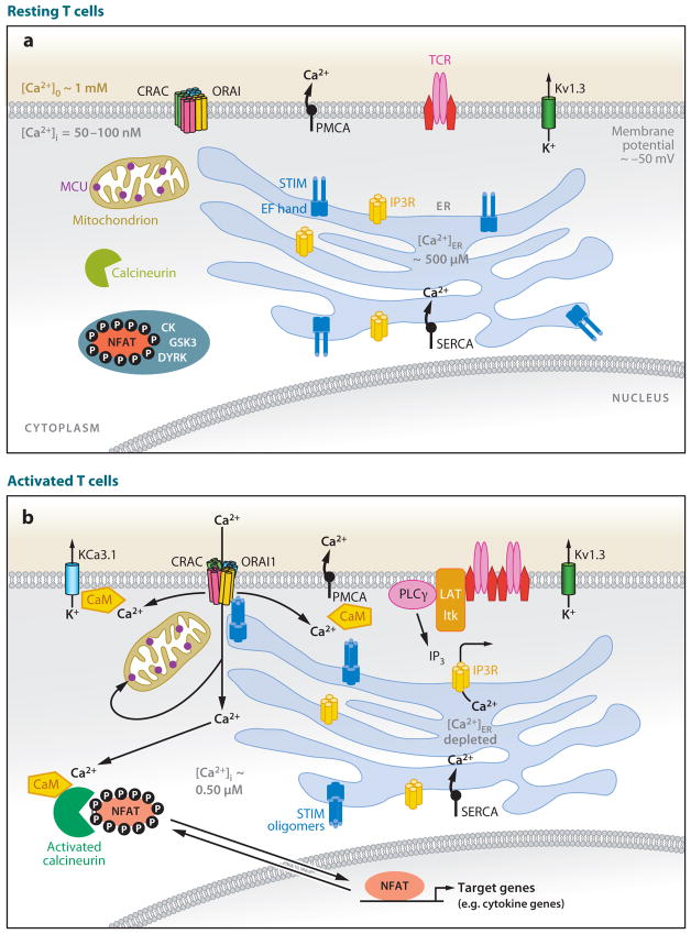
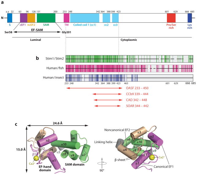
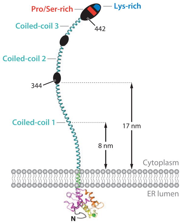
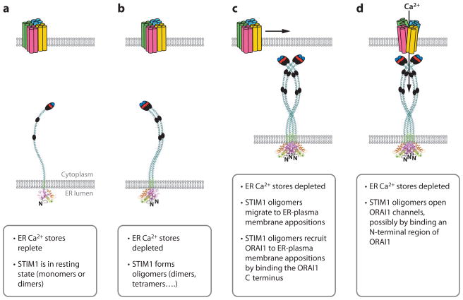
Ca(2+) entry into cells of the peripheral immune system occurs through highly Ca(2+)-selective channels known as CRAC (calcium release-activated calcium) channels. CRAC channels are a very well-characterized example of store-operated Ca(2+) channels, so designated because they open when the endoplasmic reticulum (ER) Ca(2+) store becomes depleted. Physiologically, Ca(2+) is released from the ER lumen into the cytoplasm when activated receptors couple to phospholipase C and trigger production of the second messenger inositol 1,4,5-trisphosphate (IP(3)). IP(3) binds to IP(3) receptors in the ER membrane and activates Ca(2+) release. The proteins STIM and ORAI were discovered through limited and genome-wide RNAi screens, respectively, performed in Drosophila cells and focused on identifying modulators of store-operated Ca(2+) entry. STIM1 and STIM2 sense the depletion of ER Ca(2+) stores, whereas ORAI1 is a pore subunit of the CRAC channel. In this review, we discuss selected aspects of Ca(2+) signaling in cells of the immune system, focusing on the roles of STIM and ORAI proteins in store-operated Ca(2+) entry.


Figures

Figure 1
Figure 1
Schematic diagram of the signaling pathway that connects store-operated Ca2+ entry with NFAT-dependent gene transcription in T cells. (a) Resting T cells have a membrane potential (maintained primarily by Kv1.3 K+ channels) of approximately −50 mV and intracellular free Ca2+ concentrations ([Ca2+]i) of 50–100 nM that are maintained by the plasma membrane Ca2+ ATPase (PMCA), the sarco-endoplasmic reticulum Ca2+-ATPase (SERCA) that pumps Ca2+ into the lumen of the endoplasmic reticulum (ER), and electrogenic Na+-Ca2+ exchangers (NCX, not shown). Immunoreceptors include antigen receptors on T and B cells (TCR, BCR), Fcε receptors on mast cells, or Fcγ receptors on NK cells. The concentration of free Ca2+ in the ER ([Ca2+]ER) is several hundred μM; hence the EF-hand of STIM1 is saturated with Ca2+, and STIM1 does not form higher-order oligomers (dimers are depicted, but the oligomerization state of STIM1 in resting cells is not fully defined). The transcription factor NFAT is heavily phosphorylated and localized to the cytoplasm. (b) Activated T cells. T cell receptors assemble into signaling complexes that contain scaffold proteins such as LAT and SLP-76, tyrosine kinases such as Lck, ZAP70, and Itk, and phospholipase C (PLC)γ (not all of which are shown). Inositol 1,4,5-trisphosphate (IP3) produced by PLCγbinds to IP3 receptors in the ER membrane, causing the release of Ca2+ from the ER. As a result of the depletion of ER Ca2+ stores, Ca2+ dissociates from EF-hand 1 of STIM1 and causes a conformational change (unfolding of the EF-SAM domain in the ER lumen) that leads to oligomerization (tetramers are depicted, but the oligomerization state of STIM1 in activated cells is not fully defined). The STIM oligomers move to sites of ER–plasma membrane apposition, recruit ORAI proteins to these sites, and cause CRAC channels to open. The resulting increase in [Ca2+]i causes the universal and abundant cytoplasmic Ca2+ sensor calmodulin (CaM) to bind to many channels and enzymes and modulate their activity. Among the targets of CaM are the phosphatase calcineurin, which dephosphorylates NFAT and causes its nuclear translocation, thus activating NFAT-dependent transcription; the PMCA pump whose activity is increased by CaM binding; and the KCa3.1 K+ channel that maintains membrane potential and the driving force for Ca2+ entry. Activated cells also show relocalization of mitochondria toward the plasma membrane, a process expected to maintain CRAC channel activity by diminishing Ca2+-dependent inactivation. MCU: mitochondrial Ca2+ uniporter. CK1, GSK3, DYRK: NFAT kinases.
Figure 2
Figure 2
Structure and properties of STIM1. (a) Domain structure of human STIM1 (adapted with permission from Reference 27). Shown are the signal peptide (S), the canonical EF-hand 1 (cEF1), the noncanonical EF-hand 2 (ncEF2), the SAM (sterile α-motif ) domain, the transmembrane domain (TM), three predicted coiled-coil regions (cc1, cc2, and cc3), the proline- and serine-rich region, and the lysine-rich (polybasic) region at the C terminus. The EF-SAM fragment whose structure was determined by NMR spectroscopy is indicated. The region to the left of the TM is located in the ER lumen, whereas the region to the right is located in the cytoplasm. Residue numbers at the approximate boundaries of the domains are indicated above the diagram. Coiled-coils cc1 and cc2 have long been recognized in STIM proteins (97, 98, 105) and are assigned high probability in STIM1 by COILS; the predicted coiled-coil cc3 is assigned a low probability by COILS in STIM1, but a relatively high probability in Aedes aegypti Stim and Anopheles gambiae Stim, and in STIM2 when core hydrophobic positions are weighted. The existence and precise boundaries of cc3 require experimental confirmation. (b) Sequence conservation in the STIM C-terminal region. Each horizontal black bar represents the human STIM1 sequence, with gaps introduced as necessary to maintain alignment with human STIM2, fish STIM1 orthologs, or insect Stim proteins, as indicated. Vertical green lines indicate identity of the human STIM1 residue with the residue at the corresponding position of human STIM2; vertical magenta lines indicate identity of the human STIM1 residue with residues at the corresponding position in at least four of five fish orthologs; vertical blue lines indicate identity with residues in at least two of three insect Stim proteins. Adapted from Reference . (c) Structure of the EF-SAM fragment deduced by NMR spectroscopy (adapted with permission from Reference 27). Alpha-helices are depicted as cylinders. (The canonical EF-hand 1 is magenta, the noncanonical EF-hand 2 is beige, and the SAM domain is green; the Ca2+ ion bound to EF-hand 1 is a yellow sphere.) Two views related by a 90° rotation are shown.
Figure 3
Figure 3
Schematic representation of full-length STIM1. The cytoplasmic region contains three predicted coiled-coil regions (cyan), a serine- and proline-rich region (red ), and a polybasic tail (blue). The coiled coils can span the distance, estimated to be 8 nm (109) or ~17 nm (29), that separates the ER and the plasma membrane at the junctions where STIM and ORAI accumulate upon ER Ca2+ store depletion. For an explanation of the three coiled-coil regions, see the caption to Figure 2a.
Figure 4
Figure 4
Sequence of steps in store-operated Ca2+ entry. (a) Schematic diagrams of STIM1 and ORAI1 in the resting state, when ER Ca2+ stores are replete. ORAI is depicted as a tetramer for reasons discussed in the text. STIM1 is depicted as a monomer for convenience, but its oligomerization state in resting cells is not yet fully defined. (b) STIM1 oligomerization. STIM1 forms oligomers when ER stores are depleted. Oligomers are depicted here as dimers for convenience, but their stoichiometry in activated cells is unknown. (c) STIM1 redistribution. Oligomerization of STIM1 in the ER membrane is followed by migration of STIM1 to ER–plasma membrane appositions. This redistribution involves binding of the STIM1 polybasic regions to PIP2 and PIP3 in the plasma membrane. STIM1 oligomers then recruit ORAI1 to ER–plasma membrane junctions by binding a C-terminal region of ORAI1. (d ) STIM1-ORAI1 gating. STIM1 oligomers open ORAI channels, possibly by binding to an N-terminal region of ORAI1.
Figure 5
Figure 5
Amino acid sequence of human ORAI1. Residues E106, D110, D112, D114, and E190, that when mutated affect channel properties, are shown in blue. Residues R91, A103, and L194, that when mutated to W, E, and P, respectively, are associated with human immunodeficiency, are shown in red (20, 93). Residues 65 and 74 are indicated; truncated ORAI1 proteins that begin at either residue are able to assemble and function as CRAC channels (33, 36, 150).
Figure 6
Figure 6
Pedigrees of immunodeficient patients with mutations in (a–c) ORAI1 and (d ) STIM1. (Filled symbols, patients; strike-through, deceased; ?, DNA unavailable for sequencing; dot within symbol, individual is heterozygous for the mutant allele.) Adapted with permission from References , .

Similar articles

Cited by

References

    1. Berridge MJ, Lipp P, Bootman MD. The versatility and universality of calcium signaling. Nat Rev Mol Cell Biol. 2000;1:11–21. - PubMed
    1. Berridge MJ, Bootman MD, Roderick HL. Calcium signaling: dynamics, homeostasis and remodelling. Nat Rev Mol Cell Biol. 2003;4:517–29. - PubMed
    1. Carafoli E. Calcium signaling: a tale for all seasons. Proc Natl Acad Sci USA. 2002;99:1115–22. - PMC - PubMed
    1. Clapham DE. Calcium signaling. Cell. 2007;131:1047–58. - PubMed
    1. Di Capite J, Parekh AB. CRAC channels and Ca2+ signaling in mast cells. Immunol Rev. 2009;231:45–58. - PubMed

Publication types

LinkOut - more resources