Review
. 2010 Mar 22:7:5.
doi: 10.1186/1743-8977-7-5.

Asbestos, carbon nanotubes and the pleural mesothelium: a review of the hypothesis regarding the role of long fibre retention in the parietal pleura, inflammation and mesothelioma

Affiliations
Review

Asbestos, carbon nanotubes and the pleural mesothelium: a review of the hypothesis regarding the role of long fibre retention in the parietal pleura, inflammation and mesothelioma

Ken Donaldson et al. Part Fibre Toxicol. .

Abstract

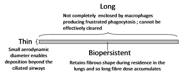
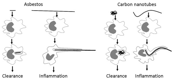
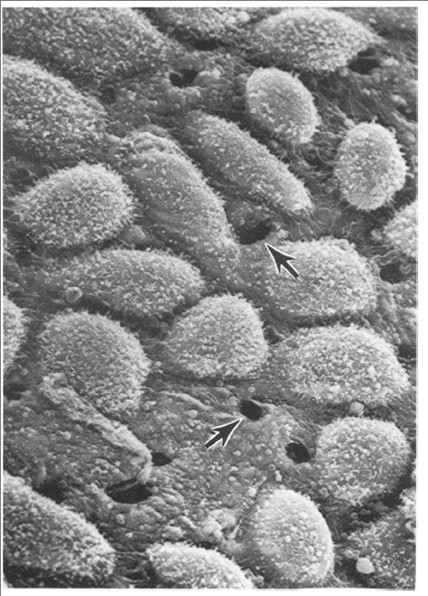
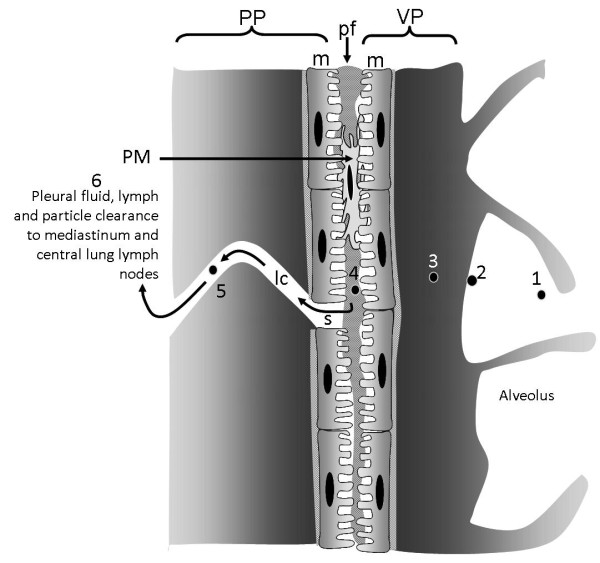
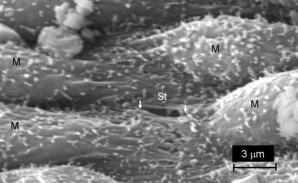
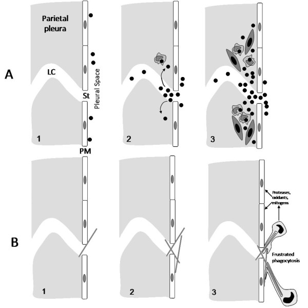
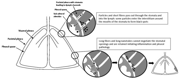
The unique hazard posed to the pleural mesothelium by asbestos has engendered concern in potential for a similar risk from high aspect ratio nanoparticles (HARN) such as carbon nanotubes. In the course of studying the potential impact of HARN on the pleura we have utilised the existing hypothesis regarding the role of the parietal pleura in the response to long fibres. This review seeks to synthesise our new data with multi-walled carbon nanotubes (CNT) with that hypothesis for the behaviour of long fibres in the lung and their retention in the parietal pleura leading to the initiation of inflammation and pleural pathology such as mesothelioma. We describe evidence that a fraction of all deposited particles reach the pleura and that a mechanism of particle clearance from the pleura exits, through stomata in the parietal pleura. We suggest that these stomata are the site of retention of long fibres which cannot negotiate them leading to inflammation and pleural pathology including mesothelioma. We cite thoracoscopic data to support the contention, as would be anticipated from the preceding, that the parietal pleura is the site of origin of pleural mesothelioma. This mechanism, if it finds support, has important implications for future research into the mesothelioma hazard from HARN and also for our current view of the origins of asbestos-initiated pleural mesothelioma and the common use of lung parenchymal asbestos fibre burden as a correlate of this tumour, which actually arises in the parietal pleura.


Figures

Figure 1
Figure 1
Diagram illustrating a pathogenic fibre according to the pathogenicity paradigm and the role of particle characteristics.
Figure 2
Figure 2
The frustrated phagocytosis paradigm as it relates to long and short fibres of asbestos (left) and various forms of carbon nanotubes (right). When confronted by short asbestos fibres or tangled, compact carbon nanotube 'particles' the macrophage can enclose them and clear them. However the macrophage cannot extend itself sufficiently to enclose long asbestos or long nanotubes, resulting in incomplete or frustrated phagocytosis, which leads to inflammation.
Figure 3
Figure 3
Frustrated phagocytosis (arrows) and the associated acute inflammatory reaction in the bronchoalveolar lavage of mice whose lungs have been instilled with long nanotubes. Aspiration of 50 μg of long fibrous multi-walled carbon nanotubes (CNT) into the lungs of C57BL/6 mice caused an acute inflammatory reaction at 24 hrs typified by a large influx of inflammatory neutrophils (PMN) into the bronchoalveolar lavage. CNT bundles and singlet fibres were seen both within macrophages (hollow arrow) and extending outside the macrophage in the process of incomplete or frustrated phagocytosis (black arrows). All images at taken at ×1000 magnification.
Figure 4
Figure 4
SEM of the surface of the peritoneal face of the diaphragm of a mouse showing the stomata (arrows) reproduced with permission from Moalli et al [4].
Figure 5
Figure 5
Multinucleate giant cell lavaged from the peritoneal cavity of a mouse instilled with long carbon nanotubes. CNT are visible in the cytoplasm (arrows); PMN = Poymorphonuclear Neutrophilic Leukocyte; L = lymphocyte (Magnification ×100).
Figure 6
Figure 6
Lesions on the peritoneal face of the diaphragm after intra-peritoneal injection of 2 forms of carbon nanotube differing in aspect ratio. The figure shows sections through the peritoneal aspect of the diaphragm of C57BL/6 mice 6 months after intra-peritoneal injection of 10 μg of two separate forms of multi-walled carbon nanotube (CNT). Sections are stained with Haematoxylin & Eosin (panels A and C) or Picro-sirius red stain which stains collagen bright red (panels B and D). Mu = Muscle of diaphragm; G = granuloma; small arrows = mesothelium; large arrows = carbon nanotubes. C and D show a large granuloma sitting atop the muscle layer, caused by the presence of CNT in the form of long fibres (open arrows). A and B show the contrasting response to CNT in the form of tightly bound dense spherical aggregates (open arrows) which produces minimal tissue reaction. All images are taken at ×100 magnification bar = 100 μm.
Figure 7
Figure 7
Diagrammatic representation of the relationship between the visceral and parietal pleurae. The visceral pleura (VP) and the parietal pleura (PP) are seen in close apposition separated by a pleural space that contains a small volume of pleural fluid (pf). Contact between the 2 pleurae is made via the mesothelial cell layers (m) on the surface of the parietal and visceral pleurae. Pleural macrophages (PM) are present in the pleural space. The rigid chest wall is tightly locked to the lungs by the adherence of the visceral pleura to the parietal pleura allowing movements of the chest wall caused by the action of the diaphragmatic muscle and intercostal muscle (IM) to expand and relax the lungs, allowing pulmonary inspiration and expiration. The pathway for particles to reach the pleural space is unknown but the path for an airborne particle (1) that deposits in the distal alveoli (2) is shown as it passes into the interstitium (3) enters the pleural space (4) and exits through a stoma in the parietal pleura (s) into a lymphatic capillary (lc, 5) to enter the lymph flow to the lymph nodes in the mediastinum and central lung.
Figure 8
Figure 8
Scanning electron micrograph image of chest wall from a normal rat showing the parietal pleural surface with mesothelial cells (M) and a stoma (white arrows, St) that is approximately 3 μm in diameter.
Figure 9
Figure 9
A) diagram showing the events leading up to formation of black spots. A1) particles enter the pleural space; A2) in focusing to exit via the stoma (St) some particles interstitialise through the loose lymphatic capillary endothelium and macrophages begin to accumulate in response; A3) macrophages and particles form a mature 'black spot' with mesenchymal cell activation and proliferation depending on the toxicity and dose of the particle. B) In B1 a single long fibre is intercepted as it attempts to negotiate the stomatal opening and is retained; 2) other fibres are caught up and there is an accumulation of retained long fibres; 3) macrophages attempt to phagocytose the fibres and undergo frustrated phagocytosis releasing a range of pro-inflammatory, genotoxic and mitogenic mediators close to the pleural mesothelial cells. PM = pleural mesothelium; St = stoma; LC = lymph capillary.
Figure 10
Figure 10
Cytospin preparations of pleural lavage cells from mice treated with short/tangled CNT (left) and long CNT (right). Arrows indicate granulocytes. Note PMN and eosinophils (arrows) indicative of inflammation in pleural leukocytes from rats exposed to the long NT only.
Figure 11
Figure 11
Histological sections through the chest wall of mice treated with long straight nanotubes (A, B) or short, tangled nanotubes (C, D) for 1 or 7 days. Note the progressive thickening of the mesothelium in A and B in response to long straight nanotubes; this contrasts with the acute thickening of the pleural layer with short/tangled CNT which shows resolution to normal mesothelium by 7 days (D). Scale bars represent 20 μm.
Figure 12
Figure 12
Hypothesised sequence of events leading to pleural responses as a consequence of long fibre retention at the parietal pleural stomatal openings.

Similar articles

Cited by

References

    1. Cugell DW, Kamp DW. Asbestos and the pleura: a review. Chest. 2004;125:1103–1117. doi: 10.1378/chest.125.3.1103. - DOI - PubMed
    1. Poland CA, Duffin R, Kinloch I, Maynard A, Wallace WA, Seaton A. Carbon nanotubes introduced into the abdominal cavity of mice show asbestos-like pathogenicity in a pilot study. Nat Nanotechnol. 2008;3:423–428. doi: 10.1038/nnano.2008.111. - DOI - PubMed
    1. Kane AB, Macdonald JL, Moalli PA. Acute injury and regeneration of mesothelial cells produced by crocidolite asbestos fibers. American Review Of Respiratory Disease. 1986;133:A198.
    1. Moalli PA, Macdonald JL, Goodglick LA, Kane AB. Acute injury and regeneration of the mesothelium in response to asbestos fibers. American Journal Of Pathology. 1987;128:426–445. - PMC - PubMed
    1. Boutin C, Dumortier P, Rey F, Viallat JR, De Vuyst P. Black spots concentrate oncogenic asbestos fibers in the parietal pleura. Thoracoscopic and mineralogic study. Am J Respir Crit Care Med. 1996;153:444–449. - PubMed

Publication types

MeSH terms