

Case Reports
doi: 10.1186/1477-7819-8-19.
Gastric glomus tumor: a case report
Affiliations
- PMID: 20307271
- PMCID: PMC2856582
- DOI: 10.1186/1477-7819-8-19
Item in Clipboard
Case Reports
Gastric glomus tumor: a case report
World J Surg Oncol.
.
Abstract
Gastric glomus tumors are rare mesenchymal tumors of the gastrointestinal tract. We describe a 72-year-old patient who presented with episodes of melena and was subsequently investigated for a tumor of the antrum of the stomach. Surgical resection revealed a 2 x 2 x 1.7 cm well circumscribed submucosal tumor, extending into the muscularis propria. The histopathologic examination of the specimen demonstrated a glomus tumor of the stomach. We discuss the preoperative investigation, the diagnostic problems and the surgical treatment of the patient with this rare submucosal lesion.
Figures
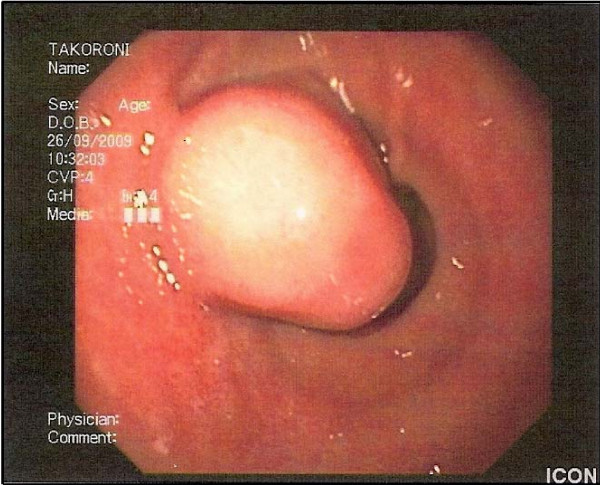
Glomus tumor of the stomach as featured on upper gastrointestinal endoscopy: a well circumscribed submucosal mass with normal overlying mucosa.
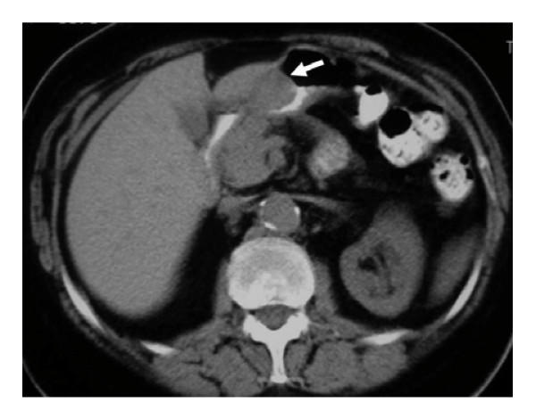
Glomus tumor of the stomach in a 72 year-old woman: unenhanced computer tomography scan shows the well-circumscribed mass (arrow) in the gastric antrum.
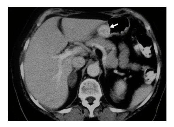
Glomus tumor of the stomach in a 72 year-old woman: On a contrast-enhanced computer tomography scan, the mass is greatly enhanced (arrow).
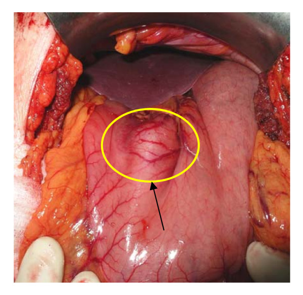
The prepyloric mass of the stomach at the lesser curvature.
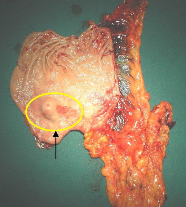
The specimen of the stomach.
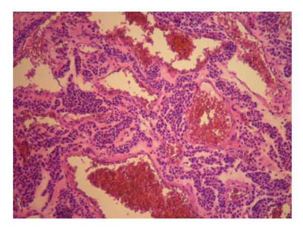
Trabeculae of tumor cells distributed around dilated and ectactic blood vessels (Hematoxylin & Eosin staining ×100).
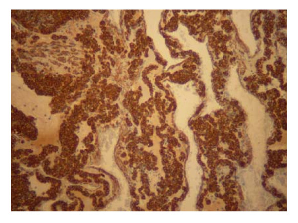
Glomus tumor of the stomach. Positive staining for smooth muscle actin (× 100).
References
-
- Kumbel JM. Glomus tumor: A benign gastric neoplasm. Mil Med. 1988;153:417–418. - PubMed
-
- Pack GT. Unusual tumors of the stomach. Ann NY Acad Sci. 1964;114:985–1011. - PubMed
-
- Enzinger FM, Weiss SW. In: Soft tissue tumors. 4. Enzinger FM, Goldblum JR, editor. St Louis, MD: Mosby; 2001. Perivascular tumors; pp. 985–1003.
Publication types
MeSH terms
LinkOut - more resources
Full Text Sources
Medical
