. 2010;12(2):R49.
doi: 10.1186/ar2960. Epub 2010 Mar 22.

Interferon-gamma inhibits interleukin-1beta-induced matrix metalloproteinase production by synovial fibroblasts and protects articular cartilage in early arthritis

Affiliations

Interferon-gamma inhibits interleukin-1beta-induced matrix metalloproteinase production by synovial fibroblasts and protects articular cartilage in early arthritis

Charlotte E Page et al. Arthritis Res Ther. 2010.

Abstract

Introduction: The first few months after symptom onset represents a pathologically distinct phase in rheumatoid arthritis (RA). We used relevant experimental models to define the pathological role of interferon-gamma (IFN-gamma) during early inflammatory arthritis.

Methods: We studied IFN-gamma's capacity to modulate interleukin-1beta (IL-1beta) induced degenerative responses using RA fibroblast-like synoviocytes (FLS), a bovine articular cartilage explant (BACE)/RA-FLS co-culture model and an experimental inflammatory arthritis model (murine antigen-induced arthritis (AIA)).

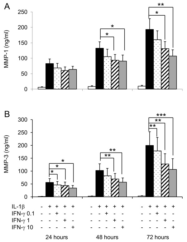
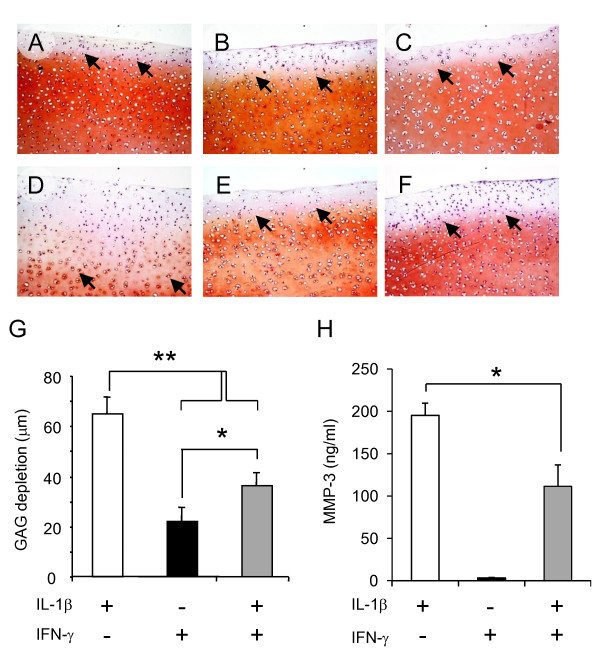
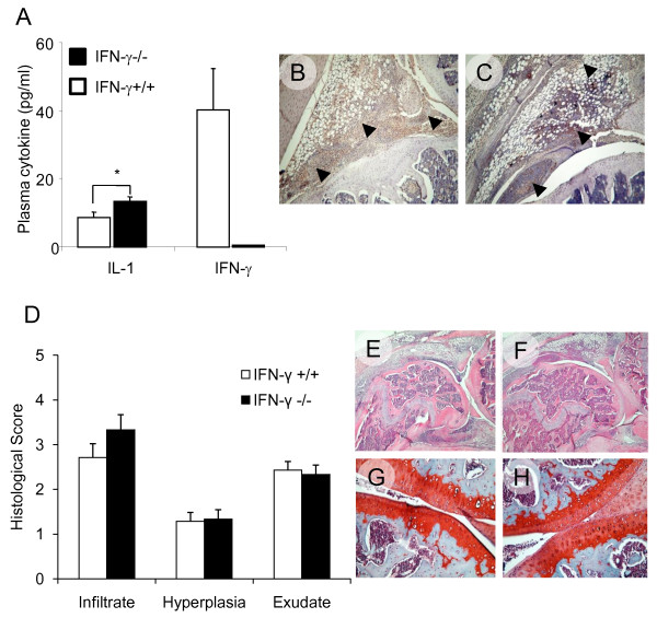
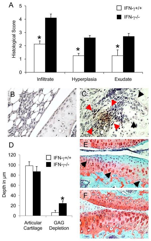
Results: IFN-gamma modulated IL-1beta driven matrix metalloproteinases (MMP) synthesis resulting in the down-regulation of MMP-1 and MMP-3 production in vitro. IFN-gamma did not affect IL-1beta induced tissue inhibitor of metalloproteinase-1 (TIMP-1) production by RA FLS but skewed the MMP/TIMP-1 balance sufficiently to attenuate glycosaminoglycan-depletion in our BACE model. IFN-gamma reduced IL-1beta expression in the arthritic joint and prevented cartilage degeneration on Day 3 of AIA.

Conclusions: Early therapeutic intervention with IFN-gamma may be critical to orchestrate tissue-protective responses during inflammatory arthritis.


Figures

Figure 1
Figure 1
IFN-γinhibits IL-1β induced MMP-1 and MMP-3 production by RA FLS in vitro. Fibroblast-like synoviocytes (FLS) were isolated from rheumatoid arthritis (RA) synovial tissue specimens. Ten RA FLS cell lines were studied in all. At fourth passage RA FLS were incubated with either culture medium, IL-1β (0.1 ng/ml), escalating doses of IFN-γ (0.1 to 10 ng/ml) or combinations of the two cytokines for 72 hours. Culture supernatants were harvested at 24, 48 and 72 hours. Protease concentrations were measured by ELISA; (A) MMP-1 and (B) MMP-3 (mean ± SEM) values are reported. Tukey's test was used in conjunction with a one-way analysis of variance to analyze differences in protease levels. P values less than 0.05 were considered statistically significant; * P < 0.05, **P < 0.01 and ***P < 0.001.
Figure 2
Figure 2
IFN-γ does not affect tissue protective TIMP-1 secretion by RA FLS in vitro. Fibroblast-like synoviocytes (FLS) were cultured from synovial tissue specimens obtained from rheumatoid arthritis (RA) patients at synovectomy or joint replacement. Eight cell lines were used at fourth passage. Cells were incubated in culture medium or in medium containing escalating doses of IFN-γ (0.1 to 10 ng/ml), IL-1β (0.1 ng/ml) or combinations of the two cytokines for 72 hours. TIMP-1 levels were quantified by ELISA in culture supernatants at 24, 48 and 72 hours; mean ± SEM TIMP-1 concentrations are reported. (A) TIMP-1 levels were significantly increased by IL-1β but not escalating concentrations of IFN-γ. IFN-γ did not affect IL-1β induced TIMP-1 production by RA FLS; (B) represents data obtained for 10 ng/ml IFN-γ ± IL-1β (0.1 ng/ml). Tukey's test was used in conjunction with a one-way analysis of variance to analyze differences in TIMP-1 levels. P values less than 0.05 were considered statistically significant. NS denotes non-significant change, *P < 0.05 and **P < 0.005.
Figure 3
Figure 3
IFN-γ protects articular cartilage from GAG depletion mediated by IL-1β activated RA FLS ex vivo. Full depth articular cartilage explants were obtained from bovine limbs (BACE). Two 12-well plates were set up in parallel, one with RA FLS the second without RA FLS. At the beginning of each experiment, one BACE was place in each well. BACE (± RA FLS) were incubated in DMEM/F12 or medium containing; 0.1 ng/ml IL-1β, 10 ng/ml IFN-γ or IL-1β with IFN-γ for 72 hours when cartilage explants and supernatants were reserved. Three RA FLS cell lines were tested; cytokines (alone or in combination) were examined in duplicate. Cartilage depletion at end point was visualised in Safranin-O/Fast Green-stained sections. Representative images from one experiment are reported (A-F), reduced intensity of red stain denotes proteoglycan loss (original magnification × 20). A-C BACE cultured without RA FLS, D-E corresponding BACE from RA FLS co-cultures which had been incubated in DMEM/F12 medium containing: 0.1 ng/ml IL-1β (A and D), 10 ng/ml IFN-γ (B and E) or IL-1β with IFN-γ (C and F) for 72 hours. The depth of GAG depletion (μm) in each BACE, measured from the articular surface to the red/orange tidemark (denoted by black block arrow) is presented graphically in (G); mean ± SEM for three experiments reported. MMP-3 was measured by ELISA in culture supernatants harvested from each well after 72 hours in culture. (H) MMP-3 (ng/ml); mean ± SEM for three experiments. One-way analysis of variance was used to analyze differences in GAG depletion and MMP-3 levels: *P < 0.05 and **P < 0.0001.
Figure 4
Figure 4
Development and progression of early arthritis in IFN-γ deficient mice. Mono-articular antigen-induced arthritis (AIA) was triggered in mice previously immunized against methylated bovine serum albumin (mBSA); arthritis was induced after an intra-articular (i.a.) injection of mBSA. Plasma and synovial cytokine expression was assessed together with histological measures of arthritis severity during early arthritis (one day after i.a. mBSA injection). (A) Plasma cytokine expression (mean ± SEM; pg/ml) shown in wild-type and IFN-γ-/- mice one day after AIA induction; *P < 0.05. (B) and (C), representative immunohistochemical staining for IL-1β in joint sections from IFN-γ+/+ and IFN-γ-/- mice respectively; intense positive brown staining (denoted by black block arrows) present in the synovial lining layer, joint exudate and focal areas of inflamed synovium, (original magnification ×10). Arthritis severity was graded in histological sections from IFN-γ+/+ (n = 7) and age matched IFN-γ-/- (n = 6) mice one day post arthritis induction. (D). Individual parameters of arthritis severity (mean ± SEM) reported. (E). Representative H&E stained para-sagittal section demonstrating histopathology of a IFN-γ+/+ knee joint processed one day post arthritis induction. (F). Corresponding section from IFN-γ-/-, showing comparable cellular infiltration, synovial hyperplasia and joint exudate (original magnification ×4). (G) and (H). Representative, Safranin-O/Fast green stained, para-sagittal section demonstrating typical cartilage architecture in arthritic knees on Day 1. Replete GAG staining (red) in articular cartilage; (G) IFN-γ+/+ and (H) IFN-γ-/- (original magnification ×20).
Figure 5
Figure 5
IFN-γ protects the synovial joint from degenerative articular changes in early experimental arthritis. Mono-articular antigen-induced arthritis (AIA) was triggered in mice previously immunized against methylated bovine serum albumin (mBSA); arthritis was induced after an intra-articular (i.a.) injection of mBSA. Changes in joint architecture were measured histologically in joint tissue sections from IFN-γ+/+ (n = 8) and age matched IFN-γ-/- (n = 10) three days after AIA induction. (A). Individual parameters of arthritis severity were determined from H&E stained arthritic knee joints; (mean ± SEM) reported; *P < 0.01. IL-1β expression was visualised in arthritic joints excised three days post AIA induction using cytokine specific immunohistochemistry. (B) and (C). Representative paraffin wax sections from IFN-γ+/+ and IFN-γ-/- stained for IL-1β (original magnification ×40). Negligible IL-1β staining observed in synovial tissues from IFN-γ+/+ mice (B). In IFN-γ-/- mice (C) intense IL-1β staining often seen in the pannus (denoted by red block arrows); articular cartilage frequently found damaged with surface fibrillation and clefts extending into transitional zone (denoted by black block arrows). Serial sections were stained with Safranin-O/Fast green. Cartilage depth and the depth of GAG depletion were calculated for each mouse from an average of five fields per femoral head. (D). Mean ± SEM depths in μm reported for IFN-γ+/+ and IFN-γ-/- (*P < 0.01). (E). Representative Safranin-O/Fast green stained para-sagittal section from IFN-γ-/- demonstrating severe GAG depletion (block arrows) and reduced cellularity. (F). Corresponding section from a IFN-γ+/+ knee joint, minimal GAG depletion observed in the cartilage on the articular surfaces (original magnification ×20).

References

    1. Ring GH, Dai Z, Saleem S, Baddoura FK, Lakkis FG. Increased susceptibility to immunologically mediated glomerulonephritis in IFN-gamma-deficient mice. J Immunol. 1999;163:2243–2248. - PubMed
    1. Jones LS, Rizzo LV, Agarwal RK, Tarrant TK, Chan CC, Wiggert B, Caspi RR. IFN-gamma-deficient mice develop experimental autoimmune uveitis in the context of a deviant effector response. J Immunol. 1997;158:5997–6005. - PubMed
    1. Ferber IA, Brocke S, Taylor-Edwards C, Ridgway W, Dinisco C, Steinman L, Dalton D, Fathman CG. Mice with a disrupted IFN-gamma gene are susceptible to the induction of experimental autoimmune encephalomyelitis (EAE) J Immunol. 1996;156:5–7. - PubMed
    1. Eriksson U, Kurrer MO, Sebald W, Brombacher F, Kopf M. Dual role of the IL-12/IFN-gamma axis in the development of autoimmune myocarditis: induction by IL-12 and protection by IFN-gamma. J Immunol. 2001;167:5464–5469. - PubMed
    1. Vermeire K, Heremans H, Vandeputte M, Huang S, Billiau A, Matthys P. Accelerated collagen-induced arthritis in IFN-gamma receptor-deficient mice. J Immunol. 1997;158:5507–5513. - PubMed

Publication types

MeSH terms