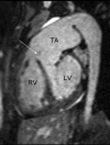
Truncus arteriosus with aortic arch interruption: cardiovascular magnetic resonance findings in the unrepaired adult
- PMID: 20307275
- PMCID: PMC2846925
- DOI: 10.1186/1532-429X-12-16
Truncus arteriosus with aortic arch interruption: cardiovascular magnetic resonance findings in the unrepaired adult
Abstract
Truncus arteriosus (TA) is a rare congenital condition defined as a single arterial vessel arising from the heart that gives origin to the systemic, pulmonary and coronary circulations. We discuss the unique case of a 28 year-old female patient with unrepaired TA and interruption of the aortic arch who underwent cardiovascular magnetic resonance (CMR).
Figures


Similar articles
-
Truncus Arteriosus With Double Aortic Arch.World J Pediatr Congenit Heart Surg. 2020 Jul;11(4):507-508. doi: 10.1177/2150135120904323. World J Pediatr Congenit Heart Surg. 2020. PMID: 32645768
-
Truncus arteriosus with double aortic arch: A rare association.Turk J Pediatr. 2017;59(2):221-223. doi: 10.24953/turkjped.2017.02.020. Turk J Pediatr. 2017. PMID: 29276881
-
Truncus arteriosus with interrupted aortic arch in very low birth weight infants.Asian Cardiovasc Thorac Ann. 2018 Sep;26(7):570-573. doi: 10.1177/0218492316647217. Epub 2016 May 5. Asian Cardiovasc Thorac Ann. 2018. PMID: 27151928
-
Repair of persistent truncus arteriosus with interrupted aortic arch.Eur J Cardiothorac Surg. 2005 Nov;28(5):736-41. doi: 10.1016/j.ejcts.2005.08.014. Epub 2005 Sep 27. Eur J Cardiothorac Surg. 2005. PMID: 16194613 Review.
-
[Truncus arteriosus communis and interrupted aortic arch].Arch Inst Cardiol Mex. 1994 Jan-Feb;64(1):57-62. Arch Inst Cardiol Mex. 1994. PMID: 8179438 Review. Spanish.
Cited by
-
An unusual combination of persistent silent truncus arteriosus Type-II with ascending aortic aneurysm.J Surg Case Rep. 2020 Jul 31;2020(7):rjaa216. doi: 10.1093/jscr/rjaa216. eCollection 2020 Jul. J Surg Case Rep. 2020. PMID: 32760491 Free PMC article.
-
Review of Journal of Cardiovascular Magnetic Resonance 2011.J Cardiovasc Magn Reson. 2012 Nov 18;14(1):78. doi: 10.1186/1532-429X-14-78. J Cardiovasc Magn Reson. 2012. PMID: 23158097 Free PMC article. Review.
-
Truncus arteriosus 5th decade transesophageal and transthoracic echocardiogram features.J Cardiol Cases. 2014 May 17;10(1):25-30. doi: 10.1016/j.jccase.2014.04.001. eCollection 2014 Jul. J Cardiol Cases. 2014. PMID: 30534217 Free PMC article.
-
Discontinuity of the arch beyond the origin of the left subclavian artery in an adult: Interruption or coarctation?Ann Pediatr Cardiol. 2018 Jan-Apr;11(1):92-96. doi: 10.4103/apc.APC_91_17. Ann Pediatr Cardiol. 2018. PMID: 29440839 Free PMC article.
-
Truncus arteriosus with persistent left superior vena cava: cardiac computed tomography findings in an unrepaired adult patient.J Clin Imaging Sci. 2013 Oct 29;3:47. doi: 10.4103/2156-7514.120787. eCollection 2013. J Clin Imaging Sci. 2013. PMID: 24228215 Free PMC article.
References
-
- Keith JD. Heart Disease in Infancy and Children. 3. New York: Macmillan; 1978. Prevalence, incidence, and epidemiology.
-
- Collett RW, Edwards JE. Persistent truncus arteriosus; a classification according to anatomic types. Surg Clin North Am. 1949;29:1245–1270. - PubMed
-
- Konstantinov IE, Karamlou T, Blackstone EH, Mosca RS, Lofland GK, Caldarone CA, Williams WG, Mackie AS, McCrindle BW. Truncus arteriosus associated with interrupted aortic arch in 50 neonates: a Congenital Heart Surgeons Society study. Ann Thorac Surg. 2006;81:214–222. doi: 10.1016/j.athoracsur.2005.06.072. - DOI - PubMed
Publication types
MeSH terms
Substances
LinkOut - more resources
Full Text Sources

