. 2010 Mar 22:7:6.
doi: 10.1186/1743-8977-7-6.

Ultrafine particles from diesel vehicle emissions at different driving cycles induce differential vascular pro-inflammatory responses: implication of chemical components and NF-kappaB signaling

Affiliations

Ultrafine particles from diesel vehicle emissions at different driving cycles induce differential vascular pro-inflammatory responses: implication of chemical components and NF-kappaB signaling

Rongsong Li et al. Part Fibre Toxicol. .

Abstract

Background: Epidemiological evidence supports the association between exposure to ambient particulate matter (PM) and cardiovascular diseases. Chronic exposure to ultrafine particles (UFP; Dp <100 nm) is reported to promote atherosclerosis in ApoE knockout mice. Atherogenesis-prone factors induce endothelial dysfunction that contributes to the initiation and progression of atherosclerosis. We previously demonstrated that UFP induced oxidative stress via c-Jun N-terminal Kinases (JNK) activation in endothelial cells. In this study, we investigated pro-inflammatory responses of human aortic endothelial cells (HAEC) exposed to UFP emitted from a diesel truck under an idling mode (UFP1) and an urban dynamometer driving schedule (UFP2), respectively. We hypothesize that UFP1 and UFP2 with distinct chemical compositions induce differential pro-inflammatory responses in endothelial cells.

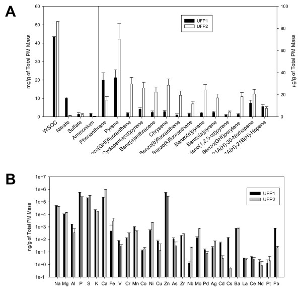
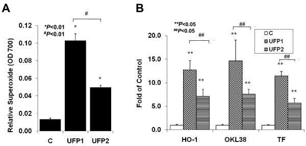
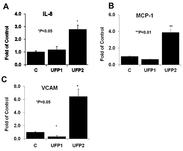
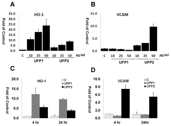
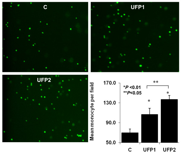
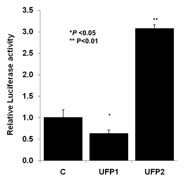
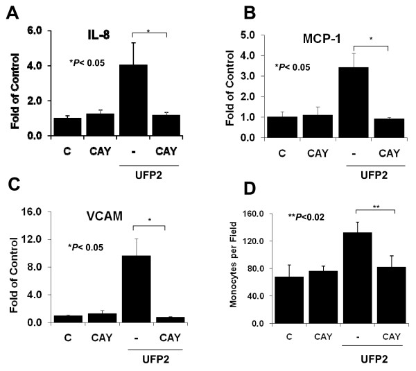
Results: UFP2 contained a higher level of redox active organic compounds and metals on a per PM mass basis than UFP1. While both UFP1 and UFP2 induced superoxide production and up-regulated stress response genes such as heme oxygenease-1 (HO-1), OKL38, and tissue factor (TF), only UFP2 induced the expression of pro-inflammatory genes such as IL-8 (2.8 +/- 0.3-fold), MCP-1 (3.9 +/- 0.4-fold), and VCAM (6.5 +/- 1.1-fold) (n = 3, P < 0.05). UFP2-exposed HAEC also bound to a higher number of monocytes than UFP1-exposed HAEC (Control = 70 +/- 7.5, UFP1 = 106.7 +/- 12.5, UFP2 = 137.0 +/- 8.0, n = 3, P < 0.05). Adenovirus NF-kappaB Luciferase reporter assays revealed that UFP2, but not UFP1, significantly induced NF-kappaB activities. NF-kappaB inhibitor, CAY10512, significantly abrogated UFP2-induced pro-inflammatory gene expression and monocyte binding.

Conclusion: While UFP1 induced higher level of oxidative stress and stress response gene expression, only UFP2, with higher levels of redox active organic compounds and metals, induced pro-inflammatory responses via NF-kappaB signaling. Thus, UFP with distinct chemical compositions caused differential response patterns in endothelial cells.


Figures

Figure 1
Figure 1
Chemical compositions of UFP1 and UFP2: (A) Comparison of PM bulk chemical species and speciated organic compounds ratios in total mass of UFP1 and UFP2. (B) Comparison of metals and trace elements ratios in total mass of UFP1 and UFP2.
Figure 2
Figure 2
UFP1 and UFP2 induced oxidative stress in HAEC. (A) UFP1 and UFP2 similarly stimulated intracellular superoxide production. HAEC were treated with 50 μg/ml (15.6 μg/cm2) of UFP1 or UFP2 for 1 hour in the presence of NBT. Superoxide production was measured as absorbance at 700 nm. (B) Both UFP1 and UFP2 stimulated the expression of oxidative stress response genes. HAEC were treated for 4 hours with 50 ug/ml of UFP1 or UFP2. The expression of stress response gene HO-1, OKL38 and TF mRNA was measured by qRT- PCR. (C = control; * versus control, n = 3, P < 0.01; ** versus control, n = 3, P < 0.05; # UFP2 versus UFP1, n = 3, P < 0.01; ## UFP2 versus UFP1, n = 3, P < 0.05)
Figure 3
Figure 3
UFP2 but not UFP1 stimulated the expression of inflammatory genes. HAEC were treated for 4 hours with 50 μg/ml (5.2 μg/cm2) of UFP1 or UFP2. The expression of inflammatory chemokine gene IL-8 (A) and MCP-1 (B) and adhesion molecule VCAM (C) was measured by qRT- PCR. (C = control; * versus control, n = 3, P < 0.05, ** versus control, P < 0.01)
Figure 4
Figure 4
Dose and time courses of UFP stimulated gene expression. (A) and (B): HAEC were treated with indicated concentration of UFP1 or UFP2 for 4 hours. The expression of HO-1 (A) and VCAM (B) was measured by qRT- PCR. (C) and (D): HAEC were treated 50 μg/ml (5.2 μg/cm2) of UFP1 or UFP2 for 4 or 24 hours. The expression of HO-1 (C) and VCAM (D) was measured by qRT- PCR. (C = control; hr = hour)
Figure 5
Figure 5
UFP1 and UFP2 stimulated monocyte binding with different capacity. HAEC grown to confluence were treated with 50 μg/ml (6.6 μg/cm2) for 5 hours. Monocyte binding assay was done with fluorescence-labeled THP-1 cells as described in methods. The averaged monocyte per high microscope field from 15 field of each condition was presented in the bar graph. (C = control; * versus control, n = 3, P < 0.01; ** UFP2 versus UFP1, n = 3, P < 0.05)
Figure 6
Figure 6
UFP2 but not UFP1 activated the NF-κB signal transduction pathway. HAEC grown to confluence were infected with Adv-LacZ and Adv-NF-κB-Luc overnight. The cells were then treated with 50 μg/ml (6.25 μg/cm2) of UFP1 or UFP2 for 24 hours. Cells were lysed and luciferase activities and β-Galactosidase activities were measured as described in methods. NF-κB reporter activities were normalized to LacZ levels and expressed as relative to control (C = control; * versus control, n = 3, P < 0.05; ** versus control, n = 3, P < 0.01)
Figure 7
Figure 7
NF-κB inhibitor CAY10512 blocked the pro-inflammatory effects of UFP2. HAEC were pretreated with 2 μg/ml of NF-κB inhibitor CAY10512 for 1 hour followed by co-treatment with 50 μg/ml (5.2 μg/cm2) of UFP2 and CAY50512 for 4 hours (gene expression) or 5 hours (monocyte binding). The expression of inflammatory gene IL-8 (A), MCP-1 (B), and VCAM (C) was measured by qRT-PCR. Monocyte binding to HAEC was measured with fluorescence-labeled THP-1 (D). (C = control; * n = 3, P < 0.05; ** n = 3, P < 0.02)

Similar articles

Cited by

References

    1. Brunekreef B, Holgate ST. Air pollution and health. Lancet. 2002;360:1233–1242. doi: 10.1016/S0140-6736(02)11274-8. - DOI - PubMed
    1. Knutsen S, Shavlik D, Chen LH, Beeson WL, Ghamsary M, Petersen F. The association between ambient particulate air pollution levels and risk of cardiopulmonary and all-cause mortality during 22 years follow-up of a non-smoking cohort. Epidemiology. 2004;15:S45–S45. doi: 10.1097/00001648-200407000-00104. - DOI
    1. Frampton MW. Systemic and cardiovascular effects of airway injury and inflammation: Ultrafine particle exposure in humans. Environmental Health Perspectives. 2001;109:529–532. doi: 10.2307/3454664. - DOI - PMC - PubMed
    1. Nemmar A, Hoet PHM, Vanquickenborne B, Dinsdale D, Thomeer M, Hoylaerts MF, Vanbilloen H, Mortelmans L, Nemery B. Passage of inhaled particles into the blood circulation in humans. Circulation. 2002;105:411–414. doi: 10.1161/hc0402.104118. - DOI - PubMed
    1. Oberdorster G. Significance of particle parameters in the evaluation of exposure-dose-response relationships of inhaled particles. Particulate Science and Technology. 1996;14:135–151. doi: 10.1080/02726359608906690. - DOI - PubMed

Publication types

MeSH terms