

Review
doi: 10.1016/j.conb.2010.02.012.
Epub 2010 Mar 20.
Function of inhibition in visual cortical processing
Affiliations
- PMID: 20307968
- PMCID: PMC3572778
- DOI: 10.1016/j.conb.2010.02.012
Item in Clipboard
Review
Function of inhibition in visual cortical processing
Curr Opin Neurobiol.
2010 Jun.
Abstract
Although sensory processing in V1 has been extensively characterized, the role of GABAergic inhibition is still not well understood. Advances in molecular biology have now removed significant barriers to the direct investigation of inhibitory processes in vivo. Recent studies have provided important insights into the influence of GABAergic inhibition on cortical processing at both the single cell level, where inhibition helps to shape cortical receptive fields, and at the network level, where inhibition is critical for generating cortical oscillations and setting network state.
2010 Elsevier Ltd. All rights reserved.
Figures
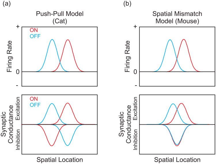
RF structure of simple cells in (a) cat and (b) mouse visual cortices. (a, Top) In cat visual cortex, simple cell RFs defined by spike rate have spatially segregated ON and OFF subregions (red and blue traces, respectively). (Bottom) This spatial segregation is also present in the RFs of excitatory and inhibitory inputs. The phase difference between the excitatory and inhibitory RFs gives rise to the classic “push-pull” antagonism. Increases in luminance over an ON subregion evoke an excitatory conductance, while the same stimulus over an OFF subregion evokes an inhibitory conductance. The opposite stimulus-response pattern is found for decreases in luminance. (b, Top) In mouse visual cortex, layer II/III simple cells also have spatially segregated ON and OFF subregions. (Bottom) This spatial segregation, however, is caused by a spatial mismatch between partially segregated ON and OFF excitatory subregions and centrally located, overlapping ON and OFF inhibitory subregions.

Neural mechanisms of spatial summation and surround suppression. (a) The spatial summation properties of cortical neurons can be quantified by plotting the response as a function of stimulus diameter. This function is dependent on stimulus contrast: the high-contrast summation field (HSF, xx trace) peaks at a smaller diameter (red arrow, preferred size) relative to the low-contrast summation field (LSF, xx trace). Surround suppression is defined as the decrease in response when the stimulus exceeds the preferred size. (b) The HSF and LSF are approximately equal in spatial extent to the spread of feed forward excitation from the LGN and the spread of intralaminar horizontal projections, respectively. The surround suppression is more consistent with cortical feedback via disynaptic inhibition. (c) The classic DOG model of surround suppression (top) predicts increased excitatory and inhibitory conductances as stimulus size is increased (bottom). (d) Experimental data, however, show that surround suppression is associated with decreases in both excitatory and inhibitory conductances.
References
-
- Moult PR. Neuronal glutamate and GABAA receptor function in health and disease. Biochem Soc Trans. 2009;37:1317–1322. - PubMed
-
- Ferster D, Miller KD. Neural mechanisms of orientation selectivity in the visual cortex. Annu Rev Neurosci. 2000;23:441–471. - PubMed
-
- Shapley R, Hawken M, Ringach DL. Dynamics of orientation selectivity in the primary visual cortex and the importance of cortical inhibition. Neuron. 2003;38:689–699. - PubMed
-
- DeAngelis GC, Robson JG, Ohzawa I, Freeman RD. Organization of suppression in receptive fields of neurons in cat visual cortex. J Neurophysiol. 1992;68:144–163. - PubMed
Publication types
MeSH terms
Substances
Grants and funding
LinkOut - more resources
Full Text Sources
