. 2010 May 21;285(21):16105-15.
doi: 10.1074/jbc.M109.081232. Epub 2010 Mar 22.

Midkine and pleiotrophin have bactericidal properties: preserved antibacterial activity in a family of heparin-binding growth factors during evolution

Affiliations

Midkine and pleiotrophin have bactericidal properties: preserved antibacterial activity in a family of heparin-binding growth factors during evolution

Sara L Svensson et al. J Biol Chem. .

Abstract

Antibacterial peptides of the innate immune system combat pathogenic microbes, but often have additional roles in promoting inflammation and as growth factors during tissue repair. Midkine (MK) and pleiotrophin (PTN) are the only two members of a family of heparin-binding growth factors. They show restricted expression during embryogenesis and are up-regulated in neoplasia. In addition, MK shows constitutive and inflammation-dependent expression in some non-transformed tissues of the adult. In the present study, we show that both MK and PTN display strong antibacterial activity, present at physiological salt concentrations. Electron microscopy of bacteria and experiments using artificial lipid bilayers suggest that MK and PTN exert their antibacterial action via a membrane disruption mechanism. The predicted structure of PTN, employing the previously solved MK structure as a template, indicates that both molecules consist of two domains, each containing three antiparallel beta-sheets. The antibacterial activity was mapped to the unordered C-terminal tails of both molecules and the last beta-sheets of the N-terminals. Analysis of the highly conserved MK and PTN orthologues from the amphibian Xenopus laevis and the fish Danio rerio suggests that they also harbor antibacterial activity in the corresponding domains. In support of an evolutionary conserved function it was found that the more distant orthologue, insect Miple2 from Drosophila melanogaster, also displays strong antibacterial activity. Taken together, the findings suggest that MK and PTN, in addition to their earlier described activities, may have previously unrealized important roles as innate antibiotics.


Figures

FIGURE 1.
FIGURE 1.
Antibacterial activity of human midkine and pleiotrophin. A–D, MK and PTN were investigated for antibacterial activity using a viable count assay. The Gram-positive pathogens S. pyogenes and S. aureus, and the Gram-negative pathogens E. coli and P. aeruginosa, were grown to mid-logarithmic phase followed by incubation with proteins at the indicated concentrations or in buffer alone, for 1 h at 37 °C. Serial dilutions were made and after culture overnight on agar plates the number of cfu were counted. The number of colonies remaining after exposure to MK and PTN, respectively, was compared with the number of cfu obtained after incubation in buffer alone, to calculate % killing. The data shown represent mean ± S.D. from three separate experiments.
FIGURE 2.
FIGURE 2.
Antibacterial activity of human MK and PTN in the presence of salt and plasma. To investigate if the presence of sodium chloride and plasma proteins interfere with the antibacterial activity of MK and PTN, respectively, the viable count assay was used. S. aureus and E. coli were grown to mid-logarithmic phase followed by incubation with MK and PTN, respectively (0.5 μm in the case of S. aureus and 1 μm in the case of E. coli) for 1 h at 37 °C. Serial dilutions were plated and the resulting cfu counted. Decrease of antibacterial activity was seen in the case of E. coli for both MK and PTN at higher concentrations of sodium chloride. Addition of plasma showed a strong inhibition of the antibacterial activity. The data shown represent mean ± S.D. from three separate experiments.
FIGURE 3.
FIGURE 3.
Membrane-disrupting properties of human MK and PTN. A and B, membrane-disruptive effects of MK (A) and PTN (B) were analyzed by measuring release of carboxyfluorescein from liposomes in the absence or presence of sodium chloride (150 mm). A dose-dependent leakage, which was not affected by the presence of sodium chloride, was seen. C, ultrastructural changes of bacterial morphology after incubation with MK or PTN was investigated using electron microscopy. S. pyogenes and E. coli were incubated in buffer alone (control), and in the presence of MK or PTN (both proteins at concentrations of 0.15 μm in the case of S. pyogenes and 0.4 μm in the case of E. coli), respectively. The samples were fixed and processed for negative staining. The electron micrographs show intact bacteria after incubation in buffer (control) while incubation in the presence of MK and PTN caused bacterial disintegration with membrane blebbing and leakage of intracellular contents. Bar, 0.5 μm.
FIGURE 4.
FIGURE 4.
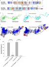
Alignment of the human MK and PTN sequences, and predicted structure of PTN. A, alignment of the MK and PTN sequences show high homology (45%) and similarity (61%). Ten cysteine residues are present, six in the N-terminal domain and four in the C-terminal domain. B, structure of MK was used as a template to predict the structure of PTN. Both molecules contain two domains consisting of three antiparallel β-sheets, held together by a hinge region. PTN has a longer unordered C-terminal tail than MK. C, models of MK and PTN showing the distribution of positively charged (blue) and negatively charged residues (red). The heparin-binding sites are indicated (circles). D, to investigate the importance of heparin-binding domains in antibacterial activity of the holoproteins, MK and PTN were preincubated with low molecular weight heparin prior to incubation with S. aureus using the viable count assay. A strong inhibition of the antibacterial activity was seen at equimolar concentrations of heparin and MK/PTN. Heparin alone did not affect the viability of bacteria. The data shown represent mean ± S.D. from three separate experiments. Statistical significance was determined based on the Student's t test for paired observations.
FIGURE 5.
FIGURE 5.
Antibacterial regions of human MK and PTN compared with orthologues from X. laevis and D. rerio. A, two regions corresponding to the highest antibacterial activity of human MK and PTN (supplemental Fig. S1) are indicated in the structural models (peptide 5: red in MK (upper) and PTN (lower); peptide 12: red in MK (upper) and in PTN (lower)). B, alignment of human MK and PTN and the corresponding orthologues from X. laevis and D. rerio. The location of peptides 5 and 12 is indicated. C, possible differences in antibacterial activity of peptides 5 and 12 was compared between species using a radial diffusion assay. Peptide 3 derived from MK and PTN, respectively, showed no activity in initial screening and thus served as negative controls. E. coli was grown to mid-logarithmic phase, dispersed in agarose that was allowed to solidify on Petri dishes. Wells were punched, and buffer alone or peptides (100 μm) were added to each well. Plates were incubated at 37 °C for 3 h to allow the peptides to diffuse. Antibacterial activity was seen as a clearing zone around each well after incubation for 18–24 h at 37 °C. Baseline value of a non-active peptide in the RDA is 0 mm. Experiments were done in triplicates, and results are shown as mean ± S.D.
FIGURE 6.
FIGURE 6.
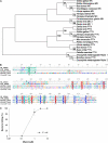
Evolution of MK and PTN. A, phylogenetic tree showing the evolutionary relationship of selected MK and PTN orthologues using the neighbor-joining method. Numbers on the branches indicate the reliability of each branch using 1,000 boot-strap replications. B, alignment of human MK and PTN sequences with Miple1 and Miple2 of D. melanogaster show complete conservation of the ten cysteine residues. Arrowheads indicate cleavage sites of signal peptides. C, antibacterial activity of Miple2 assessed by viable count assay. The number of cfu remaining after exposure to MK and PTN, respectively, were compared with the number of cfu obtained after incubation in buffer alone, to calculate % killing. The data shown represent mean ± S.D. from three separate experiments.

Similar articles

Cited by

References

    1. Zasloff M. (2002) Nature 415, 389–395 - PubMed
    1. Brogden K. A. (2005) Nat. Rev. Microbiol. 3, 238–250 - PubMed
    1. Elsbach P. (2003) J. Clin. Invest. 111, 1643–1645 - PMC - PubMed
    1. De Yang Chen Q., Schmidt A. P., Anderson G. M., Wang J. M., Wooters J., Oppenheim J. J., Chertov O. (2000) J. Exp. Med. 192, 1069–1074 - PMC - PubMed
    1. Koczulla R., von Degenfeld G., Kupatt C., Krötz F., Zahler S., Gloe T., Issbrücker K., Unterberger P., Zaiou M., Lebherz C., Karl A., Raake P., Pfosser A., Boekstegers P., Welsch U., Hiemstra P. S., Vogelmeier C., Gallo R. L., Clauss M., Bals R. (2003) J. Clin. Invest. 111, 1665–1672 - PMC - PubMed

Publication types

MeSH terms