Novel synthesis strategies for natural polymer and composite biomaterials as potential scaffolds for tissue engineering
- PMID: 20308112
- PMCID: PMC2944391
- DOI: 10.1098/rsta.2010.0009
Novel synthesis strategies for natural polymer and composite biomaterials as potential scaffolds for tissue engineering
Abstract
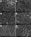
Recent developments in tissue engineering approaches frequently revolve around the use of three-dimensional scaffolds to function as the template for cellular activities to repair, rebuild and regenerate damaged or lost tissues. While there are several biomaterials to select as three-dimensional scaffolds, it is generally agreed that a biomaterial to be used in tissue engineering needs to possess certain material characteristics such as biocompatibility, suitable surface chemistry, interconnected porosity, desired mechanical properties and biodegradability. The use of naturally derived polymers as three-dimensional scaffolds has been gaining widespread attention owing to their favourable attributes of biocompatibility, low cost and ease of processing. This paper discusses the synthesis of various polysaccharide-based, naturally derived polymers, and the potential of using these biomaterials to serve as tissue engineering three-dimensional scaffolds is also evaluated. In this study, naturally derived polymers, specifically cellulose, chitosan, alginate and agarose, and their composites, are examined. Single-component scaffolds of plain cellulose, plain chitosan and plain alginate as well as composite scaffolds of cellulose-alginate, cellulose-agarose, cellulose-chitosan, chitosan-alginate and chitosan-agarose are synthesized, and their suitability as tissue engineering scaffolds is assessed. It is shown that naturally derived polymers in the form of hydrogels can be synthesized, and the lyophilization technique is used to synthesize various composites comprising these natural polymers. The composite scaffolds appear to be sponge-like after lyophilization. Scanning electron microscopy is used to demonstrate the formation of an interconnected porous network within the polymeric scaffold following lyophilization. It is also established that HeLa cells attach and proliferate well on scaffolds of cellulose, chitosan or alginate. The synthesis protocols reported in this study can therefore be used to manufacture naturally derived polymer-based scaffolds as potential biomaterials for various tissue engineering applications.
Figures


Similar articles
-
Biocompatible conducting chitosan/polypyrrole-alginate composite scaffold for bone tissue engineering.Int J Biol Macromol. 2013 Nov;62:465-71. doi: 10.1016/j.ijbiomac.2013.09.028. Epub 2013 Sep 27. Int J Biol Macromol. 2013. PMID: 24080452
-
Poly (L-lactic acid) porous scaffold-supported alginate hydrogel with improved mechanical properties and biocompatibility.Int J Artif Organs. 2016 Oct 10;39(8):435-443. doi: 10.5301/ijao.5000516. Epub 2016 Sep 3. Int J Artif Organs. 2016. PMID: 27646631
-
Chitosan-alginate hybrid scaffolds for bone tissue engineering.Biomaterials. 2005 Jun;26(18):3919-28. doi: 10.1016/j.biomaterials.2004.09.062. Biomaterials. 2005. PMID: 15626439
-
Alginate composites for bone tissue engineering: a review.Int J Biol Macromol. 2015 Jan;72:269-81. doi: 10.1016/j.ijbiomac.2014.07.008. Epub 2014 Jul 11. Int J Biol Macromol. 2015. PMID: 25020082 Review.
-
A recent study of natural hydrogels: improving mechanical properties for biomedical applications.Biomed Mater. 2025 Mar 11;20(2). doi: 10.1088/1748-605X/adb2cd. Biomed Mater. 2025. PMID: 39908671 Review.
Cited by
-
An overview to nanocellulose clinical application: Biocompatibility and opportunities in disease treatment.Regen Ther. 2023 Nov 16;24:630-641. doi: 10.1016/j.reth.2023.10.006. eCollection 2023 Dec. Regen Ther. 2023. PMID: 38034858 Free PMC article. Review.
-
New Formulations of Polysaccharide-Based Hydrogels for Drug Release and Tissue Engineering.Gels. 2015 Jan 29;1(1):3-23. doi: 10.3390/gels1010003. Gels. 2015. PMID: 30674162 Free PMC article. Review.
-
In Vivo Bone Tissue Engineering Strategies: Advances and Prospects.Polymers (Basel). 2022 Aug 8;14(15):3222. doi: 10.3390/polym14153222. Polymers (Basel). 2022. PMID: 35956735 Free PMC article. Review.
-
Resorbable Nanomatrices from Microbial Polyhydroxyalkanoates: Design Strategy and Characterization.Nanomaterials (Basel). 2022 Oct 31;12(21):3843. doi: 10.3390/nano12213843. Nanomaterials (Basel). 2022. PMID: 36364619 Free PMC article.
-
Experimental Early Stimulation of Bone Tissue Neo-Formation for Critical Size Elimination Defects in the Maxillofacial Region.Polymers (Basel). 2023 Oct 26;15(21):4232. doi: 10.3390/polym15214232. Polymers (Basel). 2023. PMID: 37959911 Free PMC article.
References
-
- Ahsan T., Bellamkonda R., Nerem R. M.2008Tissue engineering and regenerative medicine: advancing toward clinical therapies Translational approaches in tissue engineering and regenerative medicine Mao J. J., Vunjak-Novakovic A. G. Mikos, Atala A.3–16Norwood, MA: Artech House
-
- Anbergen U., Oppermann W. Elasticity and swelling behavior of chemically crosslinked cellulose ethers in aqueous systems. Polymer. 1990;31:1854–1858. doi: 10.1016/0032-3861(90)90006-K. ( ) - DOI
-
- Bonfiglio M., Jeter W. S. Immunological responses to bone. Clin. Orthop. Relat. Res. 1972;87:19–27. - PubMed
Publication types
MeSH terms
Substances
Grants and funding
LinkOut - more resources
Full Text Sources

