

Biologically active collagen-based scaffolds: advances in processing and characterization
- PMID: 20308118
- PMCID: PMC2944393
- DOI: 10.1098/rsta.2010.0015
Biologically active collagen-based scaffolds: advances in processing and characterization
Abstract
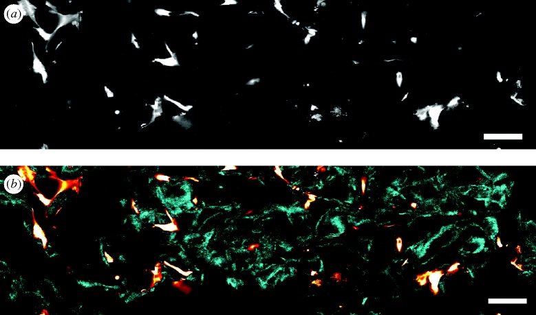
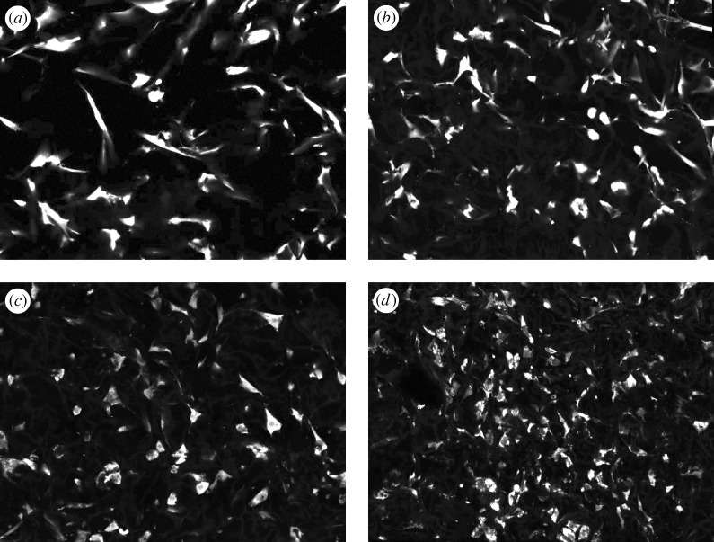
A small number of type I collagen-glycosaminoglycan scaffolds (collagen-GAG scaffolds; CGSs) have unusual biological activity consisting primarily in inducing partial regeneration of organs in the adult mammal. Two of these are currently in use in a variety of clinical settings. CGSs appear to induce regeneration by blocking the adult healing response, following trauma, consisting of wound contraction and scar formation. Several structural determinants of biological activity have been identified, including ligands for binding of fibroblasts to the collagen surface, the mean pore size (which affects ligand density) and the degradation rate (which affects the duration of the wound contraction-blocking activity by the scaffold). Processing variables that affect these determinants include the kinetics of swelling of collagen fibres in acetic acid, freezing of the collagen-GAG suspension and cross-linking of the freeze-dried scaffold. Recent developments in the processing of CGSs include fabrication of scaffolds that are paucidisperse in pore size, scaffolds with gradients in physicochemical properties (and therefore biological activity) and scaffolds that incorporate a mineral component. Advances in the characterization of the pore structure of CGSs have been made using confocal and nonlinear optical microscopy (NLOM). The mechanical behaviour of CGSs, as well as the resistance to degradative enzymes, have been studied. Following seeding with cells (typically fibroblasts), contractile forces in the range 26-450 nN per cell are generated by the cells, leading to buckling of scaffold struts. Ongoing studies of cell-seeded CGSs with NLOM have shown an advantage over the use of confocal microscopy due to the ability of the former method to image the CGS surfaces without staining (which alters its surface ligands), reduced cell photodamage, reduced fluorophore photobleaching and the ability to image deeper inside the scaffold.
Figures
Similar articles
-
Mechanical characterization of collagen-glycosaminoglycan scaffolds.Acta Biomater. 2007 Jul;3(4):463-74. doi: 10.1016/j.actbio.2006.12.009. Epub 2007 Mar 8. Acta Biomater. 2007. PMID: 17349829
-
Effect of different hydroxyapatite incorporation methods on the structural and biological properties of porous collagen scaffolds for bone repair.J Anat. 2015 Dec;227(6):732-45. doi: 10.1111/joa.12262. Epub 2014 Nov 20. J Anat. 2015. PMID: 25409684 Free PMC article.
-
Cell contraction forces in scaffolds with varying pore size and cell density.Biomaterials. 2010 Jun;31(18):4835-45. doi: 10.1016/j.biomaterials.2010.01.149. Epub 2010 Apr 2. Biomaterials. 2010. PMID: 20362329 Free PMC article.
-
Formation of collagen-glycosaminoglycan blended nanofibrous scaffolds and their biological properties.Biomacromolecules. 2005 Nov-Dec;6(6):2998-3004. doi: 10.1021/bm050318p. Biomacromolecules. 2005. PMID: 16283719
-
Surface biology of collagen scaffold explains blocking of wound contraction and regeneration of skin and peripheral nerves.Biomed Mater. 2015 Dec 23;11(1):014106. doi: 10.1088/1748-6041/11/1/014106. Biomed Mater. 2015. PMID: 26694657 Free PMC article. Review.
Cited by
-
Cell Invasion in Collagen Scaffold Architectures Characterized by Percolation Theory.Adv Healthc Mater. 2015 Jun 24;4(9):1317-21. doi: 10.1002/adhm.201500197. Epub 2015 Apr 16. Adv Healthc Mater. 2015. PMID: 25881025 Free PMC article.
-
Long-term follow-up comparison of two different bi-layer dermal substitutes in tissue regeneration: Clinical outcomes and histological findings.Int Wound J. 2020 Oct;17(5):1545-1547. doi: 10.1111/iwj.13381. Epub 2020 May 2. Int Wound J. 2020. PMID: 32359006 Free PMC article. No abstract available.
-
Reinforcement of Mono- and Bi-layer Poly(Ethylene Glycol) Hydrogels with a Fibrous Collagen Scaffold.Ann Biomed Eng. 2015 Nov;43(11):2618-29. doi: 10.1007/s10439-015-1337-0. Epub 2015 May 22. Ann Biomed Eng. 2015. PMID: 26001970 Free PMC article.
-
Bioactive IGF-1 release from collagen-GAG scaffold to enhance cartilage repair in vitro.J Mater Sci Mater Med. 2015 Jan;26(1):5325. doi: 10.1007/s10856-014-5325-y. Epub 2015 Jan 11. J Mater Sci Mater Med. 2015. PMID: 25577208 Free PMC article.
-
Preparation and characterization of elastin-like polypeptide scaffolds for local delivery of antibiotics and proteins.J Mater Sci Mater Med. 2012 Dec;23(12):2903-12. doi: 10.1007/s10856-012-4749-5. Epub 2012 Aug 28. J Mater Sci Mater Med. 2012. PMID: 22926272
References
-
- Calderwood D. A., Tuckwell D. S., Eble J., Kühn K., Humphries M. J. The integrin α1 A-domain is a binding site for collagens and laminin. J. Biol. Chem. 1997;272:12 311–12 317. - PubMed
Publication types
MeSH terms
Substances
Grants and funding
LinkOut - more resources
Full Text Sources
Other Literature Sources
