. 2010 Jul;38(13):4274-84.
doi: 10.1093/nar/gkq174. Epub 2010 Mar 22.

Quantitative profiling of the full APOBEC3 mRNA repertoire in lymphocytes and tissues: implications for HIV-1 restriction

Affiliations

Quantitative profiling of the full APOBEC3 mRNA repertoire in lymphocytes and tissues: implications for HIV-1 restriction

Eric W Refsland et al. Nucleic Acids Res. 2010 Jul.

Abstract

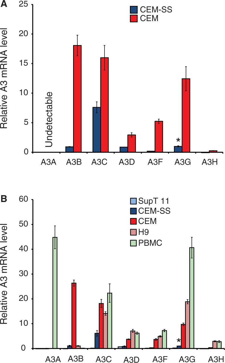
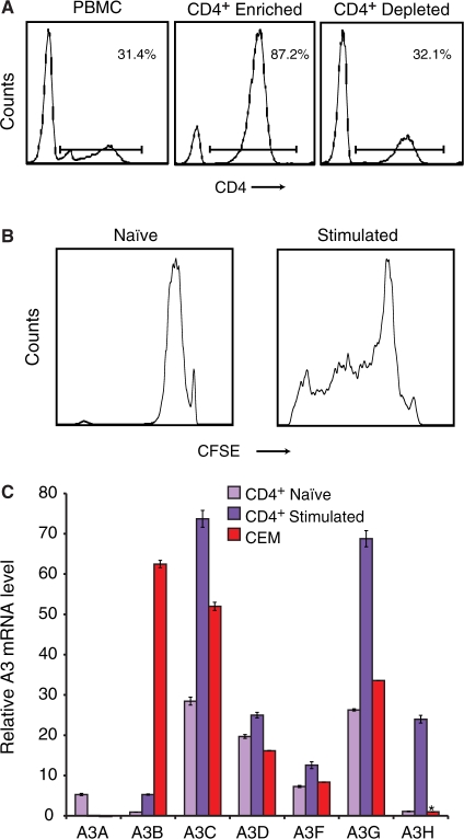
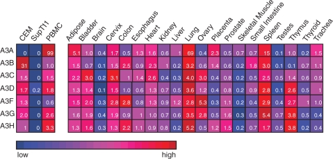
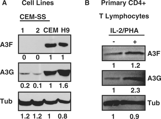
The human APOBEC3 proteins are DNA cytidine deaminases that impede the replication of many different transposons and viruses. The genes that encode APOBEC3A, APOBEC3B, APOBEC3C, APOBEC3D, APOBEC3F, APOBEC3G and APOBEC3H were generated through relatively recent recombination events. The resulting high degree of inter-relatedness has complicated the development of specific quantitative PCR assays for these genes despite considerable interest in understanding their expression profiles. Here, we describe a set of quantitative PCR assays that specifically measures the mRNA levels of each APOBEC3 gene. The specificity and sensitivity of each assay was validated using a full matrix of APOBEC3 cDNA templates. The assays were used to quantify the APOBEC3 repertoire in multiple human T-cell lines, bulk leukocytes and leukocyte subsets, and 20 different human tissues. The data demonstrate that multiple APOBEC3 genes are expressed constitutively in most types of cells and tissues, and that distinct APOBEC3 genes are induced upon T-cell activation and interferon treatment. These data help define the APOBEC3 repertoire relevant to HIV-1 restriction in T cells, and they suggest a general model in which multiple APOBEC3 proteins function together to provide a constitutive barrier to foreign genetic elements, which can be fortified by transcriptional induction.


Figures

Figure 1.
Figure 1.
A panel of quantitative PCR assays to monitor APOBEC3 mRNA levels. (A) Overview of A3 mRNA features. Each A3 mRNA is depicted to scale except 2100 nt of the A3F 3′ UTR are not shown. Inter-A3 regions of 90% or greater identity over >18 nt are highlighted in color. (B) A histogram depicting the results of running each A3 qPCR assay against each of the seven A3 cDNA control templates. (C) A graph showing the A3 qPCR assay amplification ranges and efficiencies. The mean and standard deviation of two independent experiments each consisting of three replica reactions is shown (in most instances the error is smaller than the symbol).
Figure 2.
Figure 2.
APOBEC3 expression in human T-cell lines and naïve PBMCs. (A) A3 expression in the permissive T-cell line CEM-SS and the non-permissive line CEM. Mean values and standard deviations of three independent qPCR reactions are shown for each condition. Expression is normalized to the reference gene TBP and the level of CEM-SS A3G is set to 1 to facilitate comparison (denoted by the asterisk). (B) A3 expression in the permissive T-cell lines SupT11 and CEM-SS in comparison to non-permissive lines CEM and H9. The expressed A3 repertoire in PBMCs is shown for comparison (data from an independent experiment in which CEM and CEM-SS yielded results similar to those shown here). The experimental parameters are identical to those used in panel A.
Figure 3.
Figure 3.
APOBEC3 expression in naïve and stimulated CD4+ lymphocytes. (A) Flow cytometry histograms depicting the results of CD4+ lymphocyte purification by negative selection. (B) Flow cytometry histograms of CFSE-labeled cells 4 days after mock or IL-2/PHA treatment, naïve and stimulated, respectively. (C) A3 expression in naïve and 3 day stimulated CD4+ lymphocytes. Data from CEM are shown for comparison. Expression is normalized to TBP and the level of A3H in CEM is set to 1 (denoted by the asterisk). Mean values and standard deviations of three independent qPCR reactions are shown for each condition.
Figure 4.
Figure 4.
IFN induces APOBEC3 expression in PBMCs but not CD4+ T cells. The relative A3 mRNA levels in (A) PBMCs and (B) CD4+ T lymphocytes treated for 48 h with IL-2/PHA and/or IFN. Expression is normalized to TBP and the naïve A3H level is set to 1 (denoted by the asterisk). Mean values and standard deviations of three independent qPCR reactions are shown for each condition.
Figure 5.
Figure 5.
APOBEC3 expression in human tissues. A summary of qPCR data showing the relative A3 mRNA levels in the indicated cells and tissues. The color scheme provides qualitative information, as the full range of blue (low expression) to red (high expression) color is used for each row of data. The inset numbers represent the relative levels of each A3 mRNA across the panel with the median value in each row set to 1. Since the assay efficiencies are almost identical (Figure 1C), these quantitative data can be used to compare any of the values within the table. Three replicas were done for each condition and the average values were used to construct the table (the errors were <10% and are not shown for simplicity).
Figure 6.
Figure 6.
APOBEC3 immunoblots. (A) T-cell line or (B) primary CD4+ lymphocyte (IL-2/PHA or mock treated) protein extracts were examined by immunoblotting with antibodies specific to A3F or A3G. The blots were stripped and re-probed with anti-tubulin to control for protein loading.

References

    1. Chiu YL, Greene WC. The APOBEC3 cytidine deaminases: an innate defensive network opposing exogenous retroviruses and endogenous retroelements. Annu. Rev. Immunol. 2008;26:317–353. - PubMed
    1. Goila-Gaur R, Strebel K. HIV-1 Vif, APOBEC, and intrinsic immunity. Retrovirology. 2008;5:51. - PMC - PubMed
    1. Hultquist JF, Harris RS. Leveraging APOBEC3 proteins to alter the HIV mutation rate and combat AIDS. Future Virol. 2009;4:605–619. - PMC - PubMed
    1. Malim MH, Emerman M. HIV-1 accessory proteins–ensuring viral survival in a hostile environment. Cell Host Microbe. 2008;3:388–398. - PubMed
    1. Sheehy AM, Gaddis NC, Choi JD, Malim MH. Isolation of a human gene that inhibits HIV-1 infection and is suppressed by the viral Vif protein. Nature. 2002;418:646–650. - PubMed

Publication types