. 2010 May 15;588(Pt 10):1779-90.
doi: 10.1113/jphysiol.2010.188011. Epub 2010 Mar 22.

Exercise intensity-dependent regulation of peroxisome proliferator-activated receptor coactivator-1 mRNA abundance is associated with differential activation of upstream signalling kinases in human skeletal muscle

Affiliations

Exercise intensity-dependent regulation of peroxisome proliferator-activated receptor coactivator-1 mRNA abundance is associated with differential activation of upstream signalling kinases in human skeletal muscle

Brendan Egan et al. J Physiol. .

Abstract

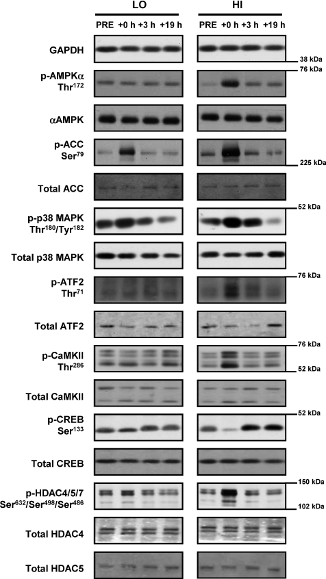
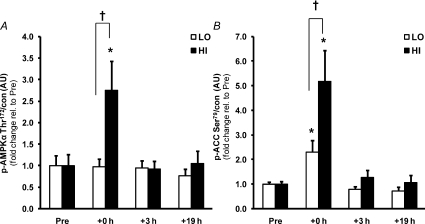
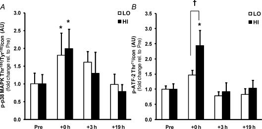
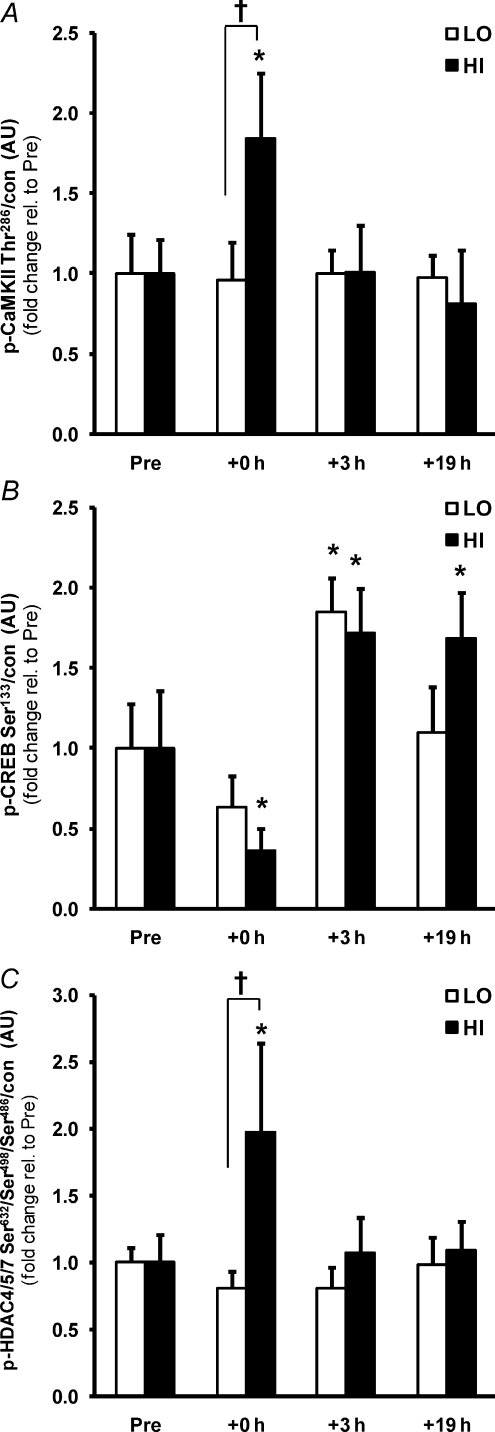
Skeletal muscle contraction increases intracellular ATP turnover, calcium flux, and mechanical stress, initiating signal transduction pathways that modulate peroxisome proliferator-activated receptor gamma coactivator-1alpha (PGC-1alpha)-dependent transcriptional programmes. The purpose of this study was to determine if the intensity of exercise regulates PGC-1alpha expression in human skeletal muscle, coincident with activation of signalling cascades known to regulate PGC-1alpha transcription. Eight sedentary males expended 400 kcal (1674 kj) during a single bout of cycle ergometer exercise on two separate occasions at either 40% (LO) or 80% (HI) of . Skeletal muscle biopsies from the m. vastus lateralis were taken at rest and at +0, +3 and +19 h after exercise. Energy expenditure during exercise was similar between trials, but the high intensity bout was shorter in duration (LO, 69.9 +/- 4.0 min; HI, 36.0 +/- 2.2 min, P < 0.05) and had a higher rate of glycogen utilization (P < 0.05). PGC-1alpha mRNA abundance increased in an intensity-dependent manner +3 h after exercise (LO, 3.8-fold; HI, 10.2-fold, P < 0.05). AMP-activated protein kinase (AMPK) (2.8-fold, P < 0.05) and calcium/calmodulin-dependent protein kinase II (CaMKII) phosphorylation (84%, P < 0.05) increased immediately after HI but not LO. p38 mitogen-activated protein kinase (MAPK) phosphorylation increased after both trials (2.0-fold, P < 0.05), but phosphorylation of the downstream transcription factor, activating transcription factor-2 (ATF-2), increased only after HI (2.4-fold, P < 0.05). Cyclic-AMP response element binding protein (CREB) phosphorylation was elevated at +3 h after both trials (80%, P < 0.05) and class IIa histone deacetylase (HDAC) phosphorylation increased only after HI (2.0-fold, P < 0.05). In conclusion, exercise intensity regulates PGC-1alpha mRNA abundance in human skeletal muscle in response to a single bout of exercise. This effect is mediated by differential activation of multiple signalling pathways, with ATF-2 and HDAC phosphorylation proposed as key intensity-dependent mediators.


Figures

Figure 6
Figure 6. Proposed model of PGC-1α gene activation in human skeletal muscle by a single bout of exercise
Acute myofibrillar contraction results in the phosphorylation (p) and activation of AMPK, CaMKII and p38 MAPK through molecular sensing of increased ATP turnover, increased calcium release from the sarcoplasmic reticulum, and mechanical stress, respectively. AMPK and CaMKII phosphorylate class IIa HDACs, leading to their nuclear exclusion and relieving the inhibitory effect of HDACs on MEF2 transcriptional activity at the MEF2 binding sequence on the PGC-1α promoter. AMPK and CaMKII also phosphorylate and activate CREB, resulting in an activating effect on the CRE sequence of the PGC-1α promoter. p38 MAPK phosphorylates and activates ATF-2, that in turn acts on the same CRE site resulting in transcriptional activation. These combined effects on the MEF2 and CRE sequences result in increased PGC-1α promoter activity and increased PGC-1α gene transcription. The intensity of the exercise bout may modulate the magnitude of this response with the phosphorylation of class IIa HDACs and ATF-2 being sensitive to the intensity of contraction, analogous to high intensity exercise (HI).
Figure 1
Figure 1. Representative immunoblots
Representative immunoblots corresponding to phosphorylated protein, total protein expression and loading control (GAPDH) measured before (Pre), immediately after (+0 h) and after 3 (+3 h) and 19 h (+19 h) of recovery from isocaloric (400 kcal) stationary cycle ergometer exercise at either 40% (low intensity, LO) or 80% (high intensity, HI) formula image
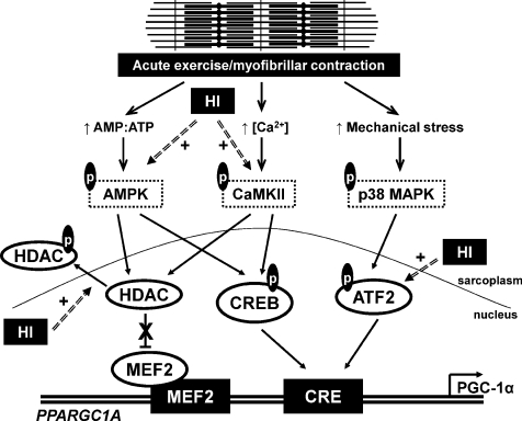
. See text for abbreviations and antibody descriptions.
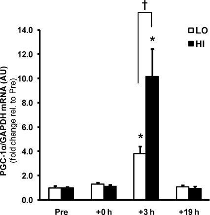
Figure 2
Figure 2. Intensity-dependent regulation of PGC-1α gene expression by a single bout of exercise
The effect of exercise intensity on PGC-1α mRNA abundance immediately after (+0 h) and during recovery (+3 h and +19 h) from isocaloric (400 kcal) exercise bouts. Open bars represent low intensity trial, LO; filled bars represent high intensity trial, HI. PGC-1α mRNA was normalized using the housekeeping gene GAPDH. Values are means ±s.e.m., n= 8. *Significantly different from baseline within same trial (P < 0.05); †significantly different from LO at same time point (P < 0.05).
Figure 3
Figure 3. Greater activation of AMPK signalling by a single bout of high intensity exercise
The effect of exercise intensity on phosphorylation of AMPK (A) and ACCβ (B) protein immediately after (+0 h) and during recovery (+3 h and +19 h) from isocaloric (400 kcal) exercise bouts. Open bars represent low intensity trial, LO; filled bars represent high intensity trial, HI. Representative immunoblots are shown in Fig. 1. Phosphorylated protein is normalised to total protein content (con) of the respective protein. Values are means ±s.e.m., n= 8. *Significantly different from baseline within same trial (P < 0.05); †significantly different from LO at same time point (P < 0.05).
Figure 4
Figure 4. Similar activation of p38 MAPK, but greater activation of ATF-2, by a single bout of high intensity compared to low intensity exercise
The effect of exercise intensity on phosphorylation of p38 MAPK (A) and ATF-2 (B) protein immediately after (+0 h) and during recovery (+3 h and +19 h) from isocaloric (400 kcal) exercise bouts. Open bars represent low intensity trial, LO; filled bars represent high intensity trial, HI. Representative immunoblots are shown in Fig. 1. Phosphorylated protein is normalised to total protein content (con) of the respective protein. Values are means ±s.e.m., n= 8. *Significantly different from baseline within same trial (P < 0.05); †significantly different from LO at same time point (P < 0.05).
Figure 5
Figure 5. Greater activation of CaMKII and HDACs, but not CREB, by a single bout of high intensity exercise
The effect of exercise intensity on phosphorylation of CaMKII (A), CREB (B) and class IIa HDAC (C) protein immediately after (+0 h) and during recovery (+3 h and +19 h) from isocaloric (400 kcal) exercise bouts. Open bars represent low intensity trial, LO; filled bars represent high intensity trial, HI. Representative immunoblots are shown in Fig. 1. Phosphorylated protein is normalised to total protein content (con) of the respective protein. Phosphorylated HDAC4/5/7 is normalised to the summed total protein content of HDAC4 and 5. Values are means ±s.e.m., n= 8. *Significantly different from baseline within same trial (P < 0.05); †significantly different from LO at same time point (P < 0.05).

Comment in

Similar articles

Cited by

References

    1. ACSM. American College of Sports Medicine Position Stand. The recommended quantity and quality of exercise for developing and maintaining cardiorespiratory and muscular fitness, and flexibility in healthy adults. Med Sci Sports Exerc. 1998;30:975–991. - PubMed
    1. Akimoto T, Li P, Yan Z. Functional interaction of regulatory factors with the Pgc-1α promoter in response to exercise by in vivo imaging. Am J Physiol Cell Physiol. 2008;295:C288–C292. - PMC - PubMed
    1. Akimoto T, Pohnert SC, Li P, Zhang M, Gumbs C, Rosenberg PB, Williams RS, Yan Z. Exercise stimulates Pgc-1α transcription in skeletal muscle through activation of the p38 MAPK pathway. J Biol Chem. 2005;280:19587–19593. - PubMed
    1. Akimoto T, Sorg BS, Yan Z. Real-time imaging of peroxisome proliferator-activated receptor-γ coactivator-1α promoter activity in skeletal muscles of living mice. Am J Physiol Cell Physiol. 2004;287:C790–C796. - PubMed
    1. Baar K. To perform your best: work hard not long. J Physiol. 2006;575:690. - PMC - PubMed

Publication types

MeSH terms

Substances