. 2010 Jun;78(6):2505-11.
doi: 10.1128/IAI.00753-09. Epub 2010 Mar 22.

Lack of interleukin-12 in p40-deficient mice leads to poor CD8+ T-cell immunity against Encephalitozoon cuniculi infection

Affiliations

Lack of interleukin-12 in p40-deficient mice leads to poor CD8+ T-cell immunity against Encephalitozoon cuniculi infection

Magali M Moretto et al. Infect Immun. 2010 Jun.

Abstract

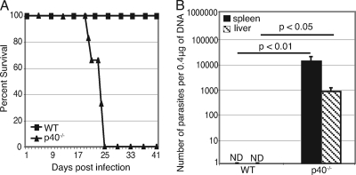
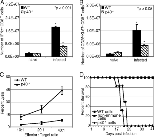
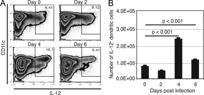
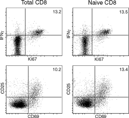
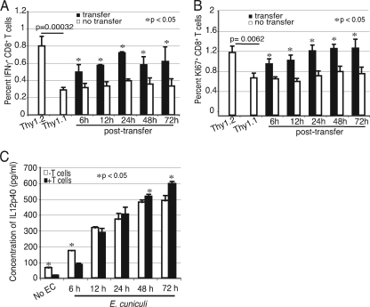
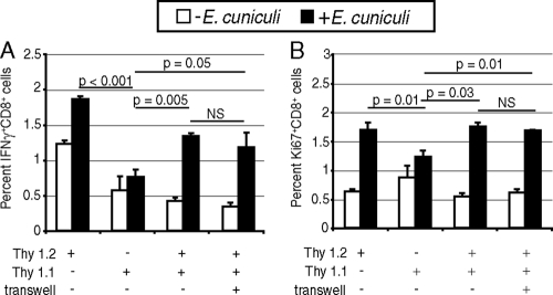
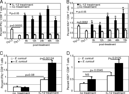
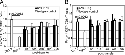
A CD8(+) T-cell response is critical for protection against Encephalitozoon cuniculi infection. However, the factors responsible for the generation of CD8(+) T-cell immunity during E. cuniculi infection and the cytokines involved in this process have not been identified. In the present study, we demonstrated that p40-deficient animals, which are unable to produce interleukin-12 (IL-12), have a serious defect in expansion of the CD8(+) T-cell response which compromises the survival of an infected host. Adoptive transfer of CD8(+) T cells from immunocompetent donors protected SCID mice infected with E. cuniculi, whereas administration of CD8(+) T cells from p40(-/-) mice failed to protect infected SCID mice. In vitro dendritic cell (DC) cultures from knockout mice pulsed with E. cuniculi spores were unable to develop a robust CD8(+) T-cell immune response. Addition of exogenous IL-12 or transfer of CD8(+) T cells that were initially primed with DC from p40(-/-) animals to DC cultures from immunocompetent mice (directly or via transwells) led to optimal expansion of these cells. This IL-12-mediated reinstatement of CD8(+) T-effector immunity was independent of gamma interferon (IFN-gamma) as addition of antibody to the cultures failed to have an effect. These studies demonstrated that IL-12 plays a predominant role in the expansion of effector CD8(+) T-cell immunity against E. cuniculi, which is critical for host survival. These findings are very important for understanding the protective immune mechanisms needed to protect an immunocompromised host against an opportunistic infection and can be extended to other microsporidial pathogens.


Figures

FIG. 1.
FIG. 1.
p40−/− mice are unable to control E. cuniculi infection. p40−/− and WT mice were infected i.p. with 107 E. cuniculi spores/mouse. (A) Animals (6 mice/group) were monitored daily for morbidity and mortality until termination of the experiment. (B) Animals (4 mice/group) were sacrificed at day 21 p.i., organs (livers and spleens) were harvested, and DNA was prepared. Parasite loads were quantified by real-time PCR. The number of parasites was determined from a standard curve after amplification from a known number of parasites. The results are representative of two experiments. ND, not determined.
FIG. 2.
FIG. 2.
Defective CD8+ T-cell response from p40−/− mice after E. cuniculi infection. p40−/− and WT mice (4 mice/group) were sacrificed at day 14 p.i., and splenocytes were prepared. (A and B) The CD8+ T-cell response was assessed by intracellular staining for IFN-γ (A) and by CD25+ KI-67+ staining (B). (C) Cytotoxicity was evaluated by the chromium release assay. CD8+ T cells were isolated by magnetic sorting at day 14 p.i., purified cells (≥92% pure) were incubated with E. cuniculi-infected radiolabeled macrophages, and cytotoxicity was measured by chromium release in the supernatants. (D) At day 14 p.i., purified CD8 T cells from spleens of WT or p40−/− mice (6 mice/group) were adoptively transferred to SCID animals (n = 6) via the i.v. route (5 × 10⁶ cells/mouse). The recipients were challenged 48 h later with 107 E. cuniculi spores. Morbidity and mortality were monitored daily until termination of the experiment. The data are representative of two experiments with similar results.
FIG. 3.
FIG. 3.
Early production of IL-12 by splenic DC after i.p. E. cuniculi infection. p40−/− and WT mice were infected i.p. with 107 E. cuniculi spores/mouse. At different time points (days 2, 4, and 6 p.i.), animals were sacrificed (3 mice/group), splenic cell suspensions were prepared, and intracellular IL-12 was measured. The results are presented as dot blots (A) and the absolute number of IL-12+ DC (B). The experiment was performed twice, and the data are representative of two experiments.
FIG. 4.
FIG. 4.
In vitro priming of total splenic CD8+ T cells or naïve CD8+ T cells by DC. Splenic DC from WT mice (4 mice/group) were isolated and incubated overnight with E. cuniculi spores. The next day, purified naïve CD8+ T cells or total splenic CD8+ T cells were added to the culture. After 72 h of incubation, cells were assessed for IFN-γ, KI67, CD25, and CD69 expression by flow cytometry. The experiment was performed twice, and the data are representative of one experiment.
FIG. 5.
FIG. 5.
p40−/− DC are unable to expand the initial CD8+ T-cell response against E. cuniculi infection. Splenic DC from WT and p40−/− mice (9 mice/group) were isolated and incubated overnight with E. cuniculi spores. The next day, CD8+ T cells from Thy1.1 and Thy1.2 animals (5 mice/group) were prepared and added to the p40−/− and WT DC cultures, respectively. At different time points following addition of T cells (6, 12, 24, 48, and 72 h), Thy1.1 CD8+ T cells were transferred from the p40−/− DC culture to a WT DC culture. At 96 h, cells were assayed for IFN-γ by intracellular staining (A) and for proliferation by using expression of the KI-67 marker (B). Supernatant from the WT DC culture was assayed for IL-12 by ELISA at different time points after addition of the T cells (6, 12, 24, 48, and 72 h) (C). Experiments were performed twice, and the results are representative of one experiment. EC, E. cuniculi.
FIG. 6.
FIG. 6.
Restoration of the CD8+ T-cell response by WT DC is contact independent. DC from WT and p40−/− animals (6 mice/group) were isolated and incubated with E. cuniculi spores as described in Materials and Methods. CD8+ T cells isolated from spleens of Thy1.1 and Thy1.2 mice (4 mice/group) were prepared and added to the p40−/− and WT DC cultures, respectively. After 6 h of incubation, Thy1.1 CD8+ T cells were transferred from the p40−/− DC culture to a WT DC culture with or without a transwell. At 96 h, the CD8+ T-cell response was assessed by detection of intracellular IFN-γ (A) and KI-67 (B). The experiment was performed twice, and the results are representative of one experiment. NS, not significant.
FIG. 7.
FIG. 7.
IL-12 treatment can restore the immune response of CD8+ T cells primed by p40−/− DC. Splenic DC from WT and p40−/− animals (9 mice/group) were incubated with E. cuniculi spores as described in the legend to Fig. 6. Affinity-purified CD8+ T cells from Thy1.1 and Thy1.2 animals (5 mice/group) were added to p40−/− and WT cultures, respectively. p40−/− DC cultures were treated with recombinant murine IL-12p70 (20 ng/ml) at different time points after addition of Thy1.1 T cells (6, 12, 24, 48, and 72 h). Thy1.2 CD8+ T cells and a WT DC culture were used as positive controls in this experiment. At 96 h, the CD8+ T-cell response was assessed by intracellular IFN-γ (A) and KI-67 (B). The nonspecific increase in the CD8+ T-cell response by IL-12 treatment alone was measured by intracellular IFN-γ (C) and KI-67 (D) expression (NS, not significant). The experiment was performed twice, and the results are representative of one experiment.
FIG. 8.
FIG. 8.
Augmentation of the IL-12-mediated CD8+ T-cell response against E. cuniculi is independent of IFN-γ. DC from WT and p40−/− animals (9 mice/group) were isolated and incubated with E. cuniculi spores. CD8+ T cells from Thy1.1 or Thy1.2 mice (5 mice/group) were added to the p40−/− or WT cultures, respectively. WT DC cultures were treated with anti-murine IFN-γ (5 μg/ml) at different time points after addition of the Thy1.2 CD8+ T cells (6, 12, 24, 48, and 72 h). Thy1.1 CD8+ T cells and a p40−/− DC culture were used as positive controls for this experiment. At 96 h, the CD8+ T-cell response was assessed by intracellular IFN-γ (A) and KI-67 (B) expression. The experiment was performed twice, and the results are representative of one experiment.

References

    1. Braunfuchsova, P., J. Salat, and J. Kopecky. 2001. CD8+ T lymphocytes protect SCID mice against Encephalitozoon cuniculi infection. Int. J. Parasitol. 31:681-686. - PubMed
    1. Braunfuchsova, P., J. Salat, and J. Kopecky. 2002. Comparison of the significance of CD4+ and CD8+ T lymphocytes in the protection of mice against Encephalitozoon cuniculi infection. J. Parasitol. 88:797-799. - PubMed
    1. Cocco, C., V. Pistoia, and I. Airoldi. 2009. New perspectives for melanoma immunotherapy: role of IL-12. Curr. Mol. Med. 9:459-469. - PubMed
    1. Curtsinger, J. M., C. M. Johnson, and M. F. Mescher. 2003. CD8 T cell clonal expansion and development of effector function require prolonged exposure to antigen, costimulation, and signal 3 cytokine. J. Immunol. 171:5165-5171. - PubMed
    1. Curtsinger, J. M., D. C. Lins, and M. F. Mescher. 2003. Signal 3 determines tolerance versus full activation of naive CD8 T cells: dissociating proliferation and development of effector function. J. Exp. Med. 197:1141-1151. - PMC - PubMed

Publication types

LinkOut - more resources