. 2010 Jun;78(6):2677-90.
doi: 10.1128/IAI.01350-09. Epub 2010 Mar 22.

Trimer stability of YadA is critical for virulence of Yersinia enterocolitica

Affiliations

Trimer stability of YadA is critical for virulence of Yersinia enterocolitica

M Schütz et al. Infect Immun. 2010 Jun.

Abstract

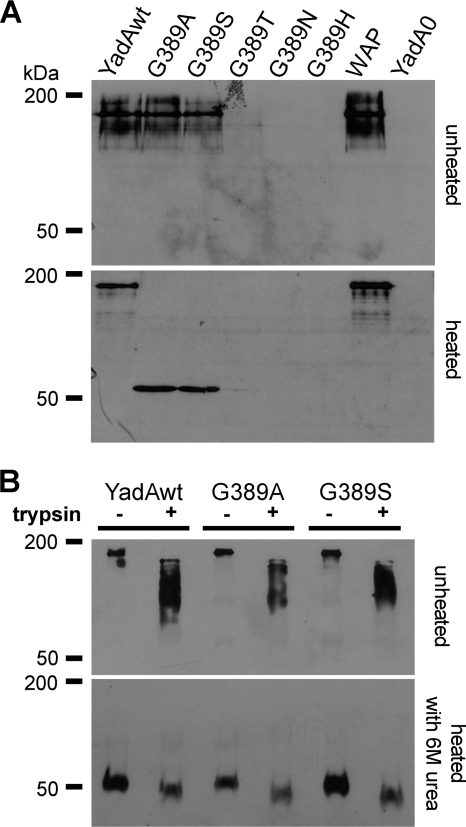
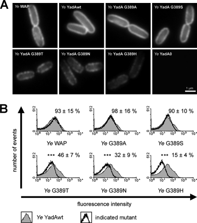
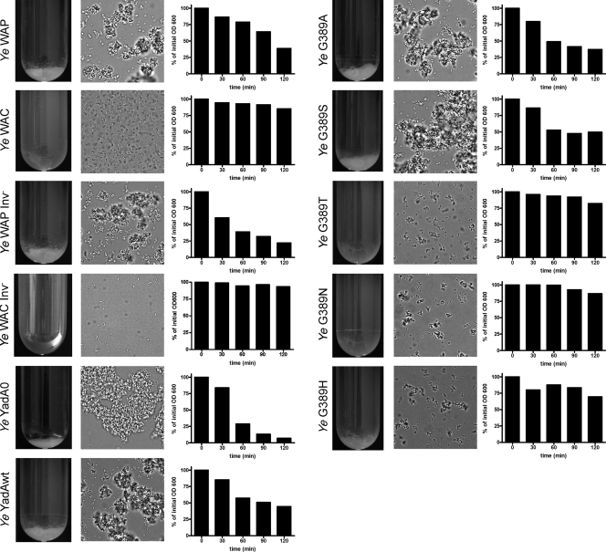
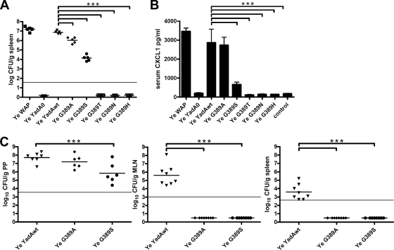
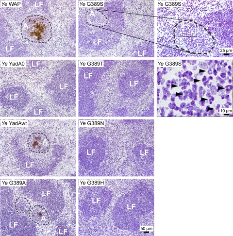
Yersinia adhesin A (YadA) is a trimeric autotransporter adhesin with multiple functions in host-pathogen interactions. The aim of this study was to dissect the virulence functions promoted by YadA in vitro and in vivo. To accomplish this, we generated Yersinia enterocolitica O:8 mutants expressing point mutations in YadA G389, a highly conserved residue in the membrane anchor of YadA, and analyzed their impact on YadA expression and virulence functions. We found that point mutations of YadA G389 led to impaired transport, stability, and surface display of YadA. YadA G389A and G389S mutants showed comparable YadA surface expression, autoagglutination, and adhesion to those of wild-type YadA but displayed reduced trimer stability and complement resistance in vitro and were 10- to 1,000-fold attenuated in experimental Y. enterocolitica infection in mice. The G389T, G389N, and G389H mutants lost trimer stability, exhibited strongly reduced surface display, autoagglutination, adhesion properties, and complement resistance, and were avirulent (>10,000-fold attenuation) in mice. Our data demonstrate that G389 is a critical residue of YadA, required for optimal trimer stability, transport, surface display, and serum resistance. We also show that stable trimeric YadA protein is essential for virulence of Y. enterocolitica.


Figures

FIG. 1.
FIG. 1.
Expression, stability, and trypsin accessibility of YadA and mutant versions expressed by Y. enterocolitica. Whole-cell lysates were prepared from Y. enterocolitica cultures grown for 2 to 3 h at 37°C. (A) Samples were either heated or not heated before separation by SDS-PAGE, and YadA was detected by Western blotting. Wild-type YadA appears as a trimer at ∼180 kDa, with monomers at 50 kDa. (B) Bacteria were grown as described above and incubated with or without 0.1 mg/ml trypsin for 1 h on ice. Afterwards, the bacterial pellet was resuspended in sample buffer. To disrupt protein trimers, samples were heated with 6 M urea to 95°C prior to loading, where indicated.
FIG. 2.
FIG. 2.
Immunofluorescence microscopy and flow cytometry analysis of Y. enterocolitica (Ye) expressing wild-type and mutant YadA. (A) Bacteria were grown for 2 h at 37°C to induce expression of YadA. YadA on the surfaces of bacteria was detected with anti-YadA antibodies and Cy2-coupled secondary antibodies. Y. enterocolitica YadA0 served as a negative control. (B) Histogram overlays of fluorescence intensity distributions in samples of bacteria expressing YadAwt or mutants. Histograms for bacteria expressing YadAwt (gray-filled area) were overlaid with the histograms for the individually indicated strains. Fluorescence of Y. enterocolitica YadA0 was also determined (not shown). The minute fluorescence of these samples was considered blank/background and subtracted from all other values. The mean fluorescence intensity of YadAwt was set to 100%. The histograms depict data for one representative experiment. Values are means with the YadA0 background subtracted ± standard deviations for three independent experiments.
FIG. 3.
FIG. 3.
Autoagglutination of Y. enterocolitica. Cultures were grown overnight at 37°C in medium containing FCS (the same results were obtained with medium without FCS [data not shown]). The tubes were then incubated at room temperature without agitation, and bacteria were allowed to settle. Photographs of culture tubes were taken after 90 min of settling. WAP, Y. enterocolitica wild-type strain; WAC, Y. enterocolitica without the virulence plasmid; WAP Inv, Y. enterocolitica wild-type strain with disrupted inv gene; WAC Inv, virulence plasmid-cured strain with disrupted inv gene. The middle columns show microscopic pictures of bacterial aggregates which were taken from the sediments in the culture tubes. The right columns show time courses of bacterial sedimentation. Optical densities of bacterial culture supernatants were determined after bacteria were allowed to settle for the indicated times.
FIG. 4.
FIG. 4.
Adhesion of Y. enterocolitica to collagen type I and HeLa cells. Y. enterocolitica strains expressing wild-type and mutant YadA were allowed to adhere to collagen-coated coverslips (A) or to HeLa cells (B). Weakly bound bacteria were washed off, and adhering bacteria were counted. Y. enterocolitica YadAwt adherence was set to 100%. The values are means ± standard deviations and are representative of three independent experiments.
FIG. 5.
FIG. 5.
Yersinia-induced IL-8 secretion and cell invasion in vitro. (A) HeLa cells were incubated with Y. enterocolitica for 1 h, and then bacteria were killed and cells were incubated for a further 6 h. IL-8 levels in the cell culture supernatants were determined by ELISA. Tumor necrosis factor alpha (TNF-α) was used as a positive control. As a negative control, cells were incubated without bacteria. Data are means and standard deviations for 2 independent experiments. (B) Uptake of bacteria into HeLa cells was analyzed by determining the number of intracellularly viable bacteria, using a gentamicin killing assay.
FIG. 6.
FIG. 6.
Serum resistance and binding of complement regulatory components factor H and C4BP are mediated by YadA. (A) Equal numbers of bacteria were incubated with 25% NHS or HIS for 1 h at 37°C and then plated on LB agar. The bacterial survival was calculated as a percentage, taking the bacterial counts obtained with HIS as 100%. (B) Binding of serum-derived factor H and C4BP to bacteria expressing or not expressing YadA was assessed by flow cytometry. Histograms for control samples (stained with primary and secondary antibodies only; light lines) were overlaid with histograms for samples specifically stained for factor H and C4BP (bold lines). The figure depicts the histograms from one representative experiment. (C) Binding of serum-derived factor H and C4BP was assessed for all strains. Bars show background-subtracted mean fluorescence intensities. Background-subtracted signal intensities for both factor H and C4BP staining for Y. enterocolitica YadA0 and the G389T, G389N, and G389H mutants sometimes reached values below zero due to variable signal noise. To make bars visible in the diagram, these values were set to 50. The data are means ± standard deviations for two independent experiments. (D) Cofactor activities of factor H bound to Y. enterocolitica WAP, YadA0, and YadAwt and the G389A mutant. The bacteria were incubated with (+) or without (−) factor H and then intensively washed. Afterwards, the bacteria were exposed to factor I and C3b. Control reactions (controls) were carried out without bacteria. C3b and the cleavage products resulting from membrane-bound factor H cofactor activity were detected with a polyclonal antibody directed against human C3b. Inactivation of C3b is indicated by the appearance of C3b α′-chain cleavage fragments of 68, 43, and 41 kDa and by reduction of the intensity of the C3b α′-chain band.
FIG. 7.
FIG. 7.
Virulence test and CXCL1 cytokine levels in sera of infected mice. (A) Numbers of bacteria (expressed as log10 CFU per gram of tissue) in the spleens of C57BL/6 mice 1 day after intravenous infection with 105 bacteria of different Yersinia strains. (B) Cytokine levels in sera of infected mice were determined by ELISA. Sera were obtained from mice that were killed 1 day after intravenous infection with 105 Y. enterocolitica cells. Values are means for at least five animal sera ± standard deviations. (C) Bacterial loads (expressed as log10 CFU per gram of tissue) in Peyer's patches (PP), mesenteric lymph nodes (MLN), and spleens after orogastric infection of BALB/c (dose of 109 CFU at 3 days postinfection) mice. ***, P < 0.001. The horizontal lines indicate the limits of detectable CFU per gram of tissue.
FIG. 8.
FIG. 8.
Immunohistochemistry of spleens of intravenously infected mice. The spleens of Y. enterocolitica-infected mice were embedded in TissueTek and shock frozen. Seven-micrometer cryosections were stained with an antibody against Y. enterocolitica and a secondary antibody coupled to peroxidase. DAB was used as a chromogenic substrate and forms a brown precipitate. Bacteria and abscesses appear in brown. Cells were counterstained with hematoxylin. LF, lymph follicle; A, abscess. Abscesses are demarcated by dashed lines. Arrowheads point to polymorphonuclear leukocytes, found in large numbers in spleens of mice infected with the Y. enterocolitica G389S mutant, despite the fact that abscess formation was not detected.

Similar articles

Cited by

References

    1. Ackermann, N., M. Tiller, G. Anding, A. Roggenkamp, and J. Heesemann. 2008. Contribution of trimeric autotransporter C-terminal domains of oligomeric coiled-coil adhesin (Oca) family members YadA, UspA1, EibA, and Hia to translocation of the YadA passenger domain and virulence of Yersinia enterocolitica. J. Bacteriol. 190:5031-5043. - PMC - PubMed
    1. Aepfelbacher, M., R. Zumbihl, K. Ruckdeschel, C. A. Jacobi, C. Barz, and J. Heesemann. 1999. The tranquilizing injection of Yersinia proteins: a pathogen's strategy to resist host defense. Biol. Chem. 380:795-802. - PubMed
    1. Autenrieth, I. B., A. Tingle, A. Reske-Kunz, and J. Heesemann. 1992. T lymphocytes mediate protection against Yersinia enterocolitica in mice: characterization of murine T-cell clones specific for Y. enterocolitica. Infect. Immun. 60:1140-1149. - PMC - PubMed
    1. Bengoechea, J. A., H. Najdenski, and M. Skurnik. 2004. Lipopolysaccharide O antigen status of Yersinia enterocolitica O:8 is essential for virulence and absence of O antigen affects the expression of other Yersinia virulence factors. Mol. Microbiol. 52:451-469. - PubMed
    1. Bernstein, H. D. 2007. Are bacterial ‘autotransporters’ really transporters? Trends Microbiol. 15:441-447. - PubMed

Publication types

MeSH terms

LinkOut - more resources