. 2010 Jun;78(6):2812-22.
doi: 10.1128/IAI.01249-09. Epub 2010 Mar 22.

Probiotic colonization of the adherent mucus layer of HT29MTXE12 cells attenuates Campylobacter jejuni virulence properties

Affiliations

Probiotic colonization of the adherent mucus layer of HT29MTXE12 cells attenuates Campylobacter jejuni virulence properties

Abofu Alemka et al. Infect Immun. 2010 Jun.

Abstract

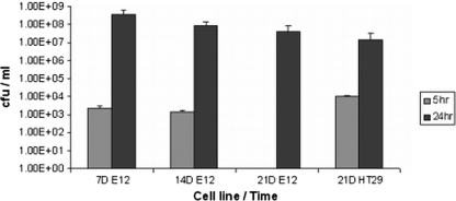
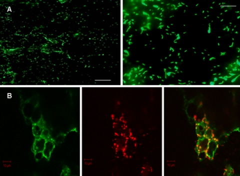
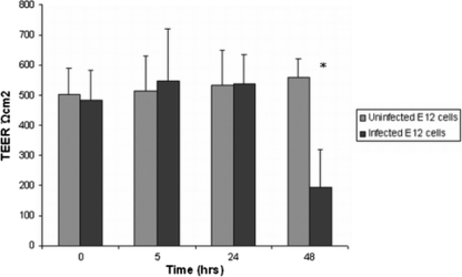
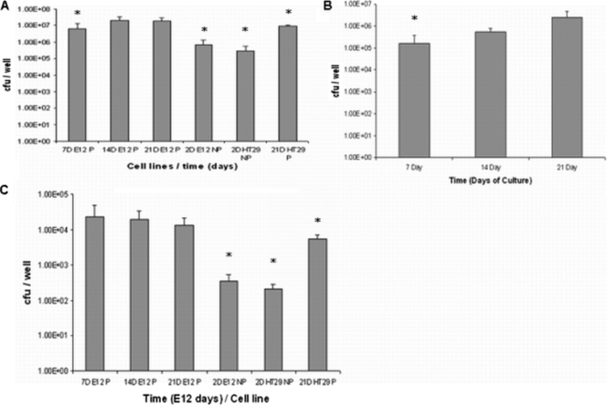
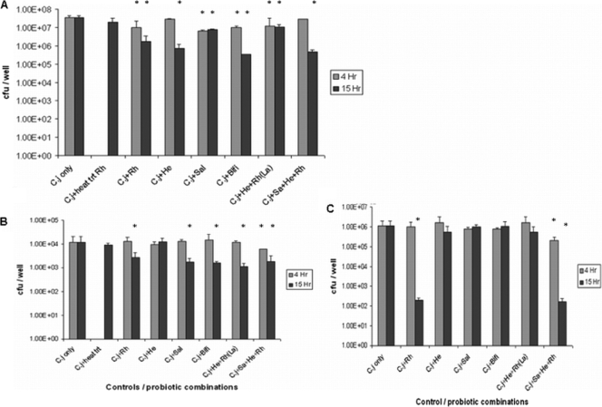
The HT29MTXE12 (E12) cell line harbors an adherent mucus layer, providing a novel technique to model mucosal infection in vitro. In this study, we have characterized the interaction of Campylobacter jejuni with the E12 cell line and exploited its unique mucus layer to examine the potential efficacy of probiotic treatment to attenuate C. jejuni virulence properties. C. jejuni 81-176 colonized and reproduced in E12 mucus. Adhesion to and internalization of C. jejuni were enhanced in E12 cells harboring mucus compared to parental cells without mucus. Translocation of C. jejuni occurred at early time points following infection. C. jejuni aligned with tight junctions and colocalized with the tight junction protein occludin, suggesting a paracellular route of translocation. Probiotic strains Lactobacillus rhamnosus R0011, Lactobacillus helveticus R0052, Lactobacillus salivarius AH102, Bifidobacterium longum AH1205, a commercial combination of L. rhamnosus R0011 and L. helveticus R0052 (Lacidofil), and a cocktail consisting of L. rhamnosus, L. helveticus, and L. salivarius (RhHeSa) colonized E12 mucus and bound to underlying cells. Probiotics attenuated C. jejuni association with and internalization into E12 cells and translocation to the basolateral medium of transwells. Live bacteria and prolonged precolonization of E12 cells with probiotics were necessary for probiotic action. These results demonstrate the potential for E12 cells as a model of mucosal pathogenesis and provide a rationale for the further investigation of probiotics as prophylaxis against human campylobacteriosis.


Figures

FIG. 1.
FIG. 1.
Interaction of C. jejuni with E12 cells. (A) Change in E12 cell mucus layer thickness and transepithelial resistance over time. The deep pink Schiff's reagent-stained mucus layer clearly increases in thickness between 7, 14, and 21 days. (i) Cells are seen adherent to the transwell filter. Remnants of liver tissue cells used for sample preparation can be visualized both above and below the filter. (ii) There is a marked increase in TEER compared to that for 21-day-cultured HT29 cells (21D HT29). Data are presented as means ± standard deviations (error bars). n = 10 for each group. Magnification, ×400. Scale bars, 150 μm. (B) Immunofluorescent micrograph of C. jejuni colonizing 21-day-cultured E12 cells. (i) Cell nuclei are visible following staining with DAPI on the transwell filter. (ii) Same field as that shown in panel Bi, showing extensive colonization by C. jejuni stained green within the mucus layer and predominantly located at the periphery of underlying cells. (iii) Some organisms (arrows) can be seen below the cell layer and above the transwell filter (translocation). Bringing organisms into focus results in some lack of definition of the filter and mucus in panels Bii and iii. Magnification, ×400. Scale bars, 10 μm. (C) C. jejuni reproduction in association with E12 cells. Three sets of 21-day-cultured E12 cells were infected with C. jejuni for 3 h. Medium was replaced with bacterium-free medium, and two sets of wells were further incubated for 24 and 48 h. A total association assay (n = 7) was performed after washes at each time point. * denotes significant difference (P < 0.05) between 24 and 48 h compared to 3 h. Error bars indicate standard deviations.
FIG. 2.
FIG. 2.
Confocal and electron micrographic images of C. jejuni on E12 cells. (A) Differential immunofluorescence distinguishes internalized (red-stained) and external adherent (yellow-green) C. jejuni organisms following infection of E12 cells. Internalized organisms commonly coalesce around cell nuclei (arrows), which are stained blue with DAPI. Magnification, ×400. Scale bar, 10 μm. Typically, some areas of the monolayer harbored mainly internalized bacteria (as seen to left of image), while in other areas both internalized and externally adherent bacteria could be visualized (right side of micrograph). (B) SEM of C. jejuni adhering to E12 cells. Organisms typically coalesced around cell-cell interfaces (yellow arrows). White arrows indicate relative effacement of microvilli on cells that are heavily colonized with C. jejuni compared to those in the upper part of the micrograph with intact microvilli and few organisms. Magnification, ×3,500. Scale bar, 2 μm. (C) SEM of two adjacent uninfected cells (slightly separated at the cell-cell junction during processing) with intact microvilli. Magnification, ×3,500. Scale bar, 2 μm.
FIG. 3.
FIG. 3.
Recovery of C. jejuni translocated to the basolateral medium after 5 h and 24 h of apical infection using 7-, 14-, and 21-day-cultured E12 cells compared to 21-day-cultured HT29 polarized cells (n = 8). Error bars indicate standard deviations. Results are presented as CFU/ml.
FIG. 4.
FIG. 4.
Pattern of C. jejuni adhesion to E12 cells. (A) Two different magnifications are shown, indicating a circular or ovoid pattern of distribution of C. jejuni as it clusters toward cell-cell junctions. Scale bars, 10 μm (left) and 5 μm (right). Magnification, ×200 (left) and ×400 (right). (B) Confocal microscopy of the tight junction protein occludin and C. jejuni. There is clear pericellular distribution of C. jejuni organisms (stained red) and colocalization with green-stained occludin protein (yellow spots). Magnification, ×630. Scale bars, 10 μm.
FIG. 5.
FIG. 5.
Transepithelial resistance values of 21-day E12 cells following C. jejuni infection remain unaffected despite very high C. jejuni translocation efficiency at 24 h. There is no evidence of loss of TEER until later time points (48 h). * denotes significant difference (P < 0.00005) compared to 48 h uninfected E12 cells (n = 12). Error bars indicate standard deviations.
FIG. 6.
FIG. 6.
Colonization, adhesion, and invasion of E12 cells. (A) C. jejuni total association (mucus-colonized, adherent, and invaded organisms) to polarized E12 cells compared to parental cell lines. Day 7-, 14-, and 21-day-cultured E12 cells are indicated as 7DE12P, 14DE12P, and 21DE12P, respectively. 2DE12NP, 2-day-cultured nonpolarized E12 cells; 2DHT29NP, 2-day-cultured nonpolarized HT29 cells; 21D HT29P, 21-day-cultured polarized HT29 cells (no mucus layer). * denotes significant difference compared to 21DE12P. 7DE12P, P < 0.005; 2DE12NP, P < 0.005; 2DHT29NP, P < 0.0005; 21DHT29P, P < 0.05 (n = 11). Error bars indicate standard deviations. (B) Adhesion of C. jejuni to E12 cells after removal of the mucus layer. * denotes significant difference compared to 14- and 21-day-cultured E12 cells. 14-day E12, P < 0.05; 21 -day E12, P < 0.05 (n = 8). Error bars indicate standard deviations. (C) C. jejuni internalization into polarized E12 cells compared to parental cell lines. * denotes significant difference compared to 21DE12P. 2DE12NP, P < 0.005; 2DHT29 NP, P < 0.05; 21DHT29P, P < 0.05 (n = 8). Error bars indicate standard deviations.
FIG. 7.
FIG. 7.
Effect of probiotics and probiotic cocktails on C. jejuni total association to (A), invasion of (B), and translocation across (C) 14-day-cultured E12 cells. (A) E12 cells were precolonized with probiotics and probiotic combinations for 4 and 15 h prior to infection with C. jejuni (C.j) for a further 24 h. Rh, L. rhamnosus; heat trt Rh, heat-treated Rh; He, L. helveticus; Sal, L. salivarius; Bifi, B. longum; La, L. rhamnosus and L. helveticus. Error bars indicate standard deviations. * denotes significant difference compared to C. jejuni only (n = 9). P < 0.005 for all groups. (B) E12 cells were precolonized with probiotics for 4 and 15 h prior to infection with C. jejuni for a further 24 h. The gentamicin protection assay was used to quantify internalized C. jejuni CFU. * denotes statistical difference compared to C. jejuni only (n = 8). P < 0.05 for all groups. Error bars indicate standard deviations. (C) E12 cells cultured for 14 days were precolonized with probiotics for 4 and 15 h prior to infection with C. jejuni for a further 24 h (n = 8). Serial dilutions of the basolateral medium were made in PBS and plated onto solid medium. Error bars indicate standard deviations. * denotes statistical difference compared to C. jejuni only. Rh, P < 0.05; 4 h Sa+He+Rh, P < 0.05; 15 h Sa+He+Rh, P < 0.05.

Similar articles

Cited by

References

    1. Altenhoefer, A., S. Oswald, U. Sonnenborn, C. Enders, J. Schulze, J. Hacker, and T. A. Oelschlaeger. 2004. The probiotic Escherichia coli strain Nissle 1917 interferes with invasion of human intestinal epithelial cells by different enteroinvasive bacterial pathogens. FEMS Immunol. Med. Microbiol. 40:223-229. - PubMed
    1. Barragan, A., F. Brossier, and L. D. Sibley. 2005. Transepithelial migration of Toxoplasma gondii involves an interaction of intercellular adhesion molecule 1 (ICAM-1) with the parasite adhesin MIC2. Cell Microbiol. 7:561-568. - PubMed
    1. Behrens, I., P. Stenberg, P. Artursson, and T. Kissel. 2001. Transport of lipophilic drug molecules in a new mucus-secreting cell culture model based on HT29-MTX cells. Pharm. Res. 18:1138-1145. - PubMed
    1. Beltinger, J., J. del Buono, M. M. Skelly, J. Thornley, R. C. Spiller, W. A. Stack, and C. J. Hawkey. 2008. Disruption of colonic barrier function and induction of mediator release by strains of Campylobacter jejuni that invade epithelial cells. World J. Gastroenterol. 14:7345-7352. - PMC - PubMed
    1. Bras, A. M., and J. M. Ketley. 1999. Transcellular translocation of Campylobacter jejuni across human polarised epithelial monolayers. FEMS Microbiol. Lett. 179:209-215. - PubMed

Publication types

LinkOut - more resources