. 2010 Apr 12;207(4):793-806.
doi: 10.1084/jem.20092545. Epub 2010 Mar 22.

Antagonism of the complement component C4 by flavivirus nonstructural protein NS1

Affiliations

Antagonism of the complement component C4 by flavivirus nonstructural protein NS1

Panisadee Avirutnan et al. J Exp Med. .

Abstract

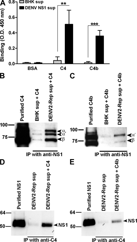
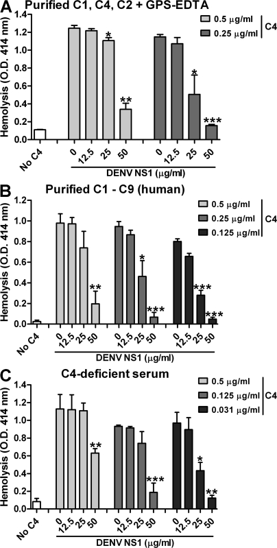
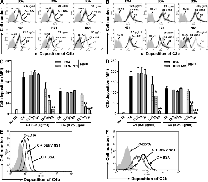
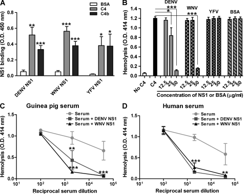
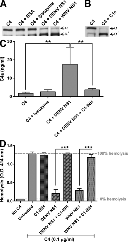
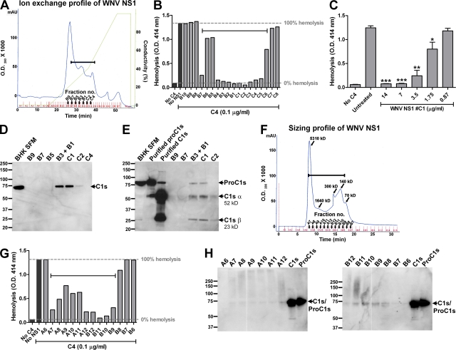
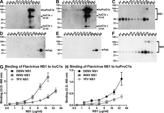
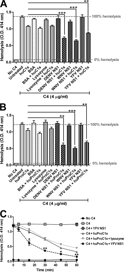
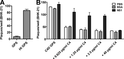
The complement system plays an essential protective role in the initial defense against many microorganisms. Flavivirus NS1 is a secreted nonstructural glycoprotein that accumulates in blood, is displayed on the surface of infected cells, and has been hypothesized to have immune evasion functions. Herein, we demonstrate that dengue virus (DENV), West Nile virus (WNV), and yellow fever virus (YFV) NS1 attenuate classical and lectin pathway activation by directly interacting with C4. Binding of NS1 to C4 reduced C4b deposition and C3 convertase (C4b2a) activity. Although NS1 bound C4b, it lacked intrinsic cofactor activity to degrade C4b, and did not block C3 convertase formation or accelerate decay of the C3 and C5 convertases. Instead, NS1 enhanced C4 cleavage by recruiting and activating the complement-specific protease C1s. By binding C1s and C4 in a complex, NS1 promotes efficient degradation of C4 to C4b. Through this mechanism, NS1 protects DENV from complement-dependent neutralization in solution. These studies define a novel immune evasion mechanism for restricting complement control of microbial infection.


Figures

Figure 1.
Figure 1.
Direct binding of DENV NS1 to C4 and C4b. (A) ELISA. Microtiter plates were coated with BSA, C4, or C4b (5 µg/ml). After incubation with serum-free supernatants from BHK-DENV2-Rep cells or control BHK cells, bound DENV NS1 was detected using DENV NS1–specific mAbs. Data are the mean ± SD for three independent experiments. Asterisks denote NS1 binding that is statistically different from binding to background (**, P < 0.005; ***, P < 0.0005). (B–E) Reciprocal co-immunoprecipitation. Serum-free supernatants from BHK-DENV2-Rep or control BHK cells were incubated with purified C4 (B and D) or C4b (C and E; 20 µg/ml) and Western blots were performed after immunoprecipitation with anti-DENV NS1 mAb (2G6)–Sepharose (B and C) or anti–human C4–Sepharose (D and E). Immunoprecipitated material was probed with a goat polyclonal anti–human C4 Ab or DENV NS1–specific mAb (1F11), respectively. Subunits and cleavage fragments of C4 and C4b are labeled to the right of the gel; a 93-kD α chain of C4; 84-kD α′ chain of C4b; and 78-kD β chain of C4 and C4b. Notably, in the starting material of this C4 preparation, 20–30% was C4b. Western blot results are representative of two or three independent experiments.
Figure 2.
Figure 2.
DENV NS1 inhibits C4 to prevent CP dependent hemolysis. (A and B) Human C4 was incubated in the presence or absence of DENV NS1 and then added to sensitized EA bearing C1 (EAC1). After three washes, the EAC14b cells were incubated with purified C2, washed, and incubated with GPS treated with EDTA (A) or purified human components (B; C3-C9) at 37°C for 30 min. After centrifugation, the supernatant was analyzed by spectrophotometry at 414 nm. (C) Single-step hemolysis assay. EA were incubated with an 80-fold dilution of C4-deficient GPS that was supplemented with human C4, with or without preincubation with DENV NS1. The data represent the mean ± SD for three independent experiments. Asterisks denote hemolysis conditions that are statistically different from untreated cells (*, P < 0.05; **, P < 0.005; ***, P < 0.0005).
Figure 3.
Figure 3.
DENV NS1 reduces C4b and C3b deposition on the surface of cells. Human C4 was incubated with serially diluted purified DENV NS1 or BSA (12.5, 25, or 50 µg/ml) for 1 h before adding EAC1. After a 15-min incubation at 30°C, EAC14b were sequentially incubated with C2 and C3. After three washes, the deposition of C4b (A and C) and C3b (B and D) was determined by incubating EAC14b2a3b with anti-C4c or anti-C3c mAb, and analyzed by flow cytometry. Examples of histogram profiles in which DENV NS1, but not BSA, blocks C4b (A) and C3b (B) deposition are depicted. In C and D, data are expressed as the specific mean fluorescence intensity (MFI) of staining. The error bars indicate SD corresponding to three independent experiments. Asterisks denote C4b or C3b deposition that is statistically different from untreated cells (*, P < 0.05; **, P < 0.005; ***, P < 0.0005). (E and F) Soluble DENV NS1 restricts complement deposition on the surface of nucleated cells. CHO cells were sensitized with rabbit anti-CHO surface antigen IgG before incubation with the mixture of DENV NS1 or BSA (25 µg/ml) and purified human CP complement components C1, C4, C2, and C3, followed by immunostaining with murine mAb to human C4d (E) and C3d (F) and analyzed by flow cytometry. EDTA (20 mM) was added to inhibit complement activation for negative controls. Examples of histogram profiles from three independent experiments in which DENV NS1, but not BSA, restricted C4b (E) and C3b (F) deposition are shown.
Figure 4.
Figure 4.
DENV and WNV NS1 inhibit CP activation via a direct interaction with C4. (A) DENV, WNV, and YFV NS1 interact with human C4/C4b. Microtiter plates were coated with BSA, C4, or C4b (5 µg/ml). After incubation with purified DENV, WNV, or YFV NS1 (5 µg/ml), bound NS1 was detected using DENV, WNV, or YFV NS1-specific mAbs. (B) EA were incubated with a 160-fold dilution of C4-deficient GPS that was supplemented with purified human C4 (63 ng/ml), with or without preincubation with DENV, WNV, or YFV NS1 or BSA (12.5–50 µg/ml). (C and D) DENV and WNV NS1 inhibit C4-mediated CP in guinea pig and human sera. EA were incubated with a 160-fold dilution of C4-deficient GPS that was supplemented with the indicated dilutions of fresh guinea pig (C) or human serum (D), with or without preincubation with DENV or WNV NS1 (50 µg/ml). Data are the mean ± SD for three independent experiments. Asterisks denote NS1 binding that is statistically different from binding to background (A) or hemolysis conditions which are statistically different from untreated cells (B–D; *, P < 0.05; **, P < 0.005; ***, P < 0.0005).
Figure 5.
Figure 5.
Purified DENV NS1 and WNV NS1 promote C4 cleavage. (A and B) Western blot analysis. (A) Purified DENV NS1, WNV NS1, BSA, or lysozyme (50 µg/ml) was preincubated with C4 (6.25 µg/ml) for 1 h on ice. Subsequently, the NS1-C4, BSA-C4, and lysozyme-C4 solutions were incubated at 30°C for 15 min. (B) Purified C4 was incubated with C1s (12 µg/ml) for 1 h at 30°C. Subunits and cleavage fragments of C4 and C4b are labeled to the right of the gel; 93 kD α chain of C4; 84 kD α′ chain of C4b. (C and D) Inhibition of C4 cleavage in the presence of NS1 by C1 inhibitor (C1-INH). (C) C4a ELISA. C4 was incubated with purified DENV NS1 or lysozyme (30 µg/ml) in the presence or absence of C1-INH (1.4 µg/ml) on ice for 1 h. (D) Single-step hemolysis assay. EA were incubated with a 160-fold dilution of C4-deficient GPS supplemented with human C4 (0.1 µg/ml) that was preincubated with DENV or WNV NS1 (30 µg/ml) in the presence or absence of C1-INH (40 µg/ml). In some experiments, C1 inhibitor (C1-INH; 40 µg/ml) was added to before the incubation with EA- and C4-deficient GPS. Data are the mean ± SD for four independent experiments. Asterisks denote C4a liberation (C) as measured by ELISA that is statistically different (**, P < 0.005) or hemolysis conditions (D) that are statistically different from untreated cells (***, P < 0.0005).
Figure 6.
Figure 6.
C1s/proC1s copurifies with NS1. (A) Two-step purification of WNV NS1. Immunoaffinity-purified WNV NS1 was passed over an ion-exchange column, and NS1 was eluted with a linear NaCl gradient (diagonal line). Silver stain and Western blot of each fraction were performed (Fig. S5 and not depicted). All fractions were tested for C4 inhibitory activity in a single-step hemolysis assay (B). C4 (0.1 µg/ml) was preincubated with an individual concentrated fraction (25 µl) before the addition of EA- and C4-deficient GPS (1:160). Fractions with C4 inhibitory activity are bracketed (A and B). (C) WNV NS1 fraction #C1 from ion exchange chromatography was twofold serially diluted and subjected to a single-step C4 hemolysis assay. (D and E) Western blot analysis for C1s/proC1s from ion exchange fractions shown in A. (D) Nonreduced SDS-PAGE. Lane 1, 100-fold concentrated serum-free culture supernatants (SFM) of BHK cells; lanes 2–7, 10-fold concentrated purified WNV NS1 fractions after ion-exchange chromatography. The proteins were separated on a 4–12% gradient nonreduced SDS-PAGE, transferred, and probed with a goat anti–human C1s serum. The anti–human C1s Ab cross-reacts with hamster C1s. (E) SDS-PAGE, reducing conditions. Lane 1, 100-fold concentrated SFM of BHK cells; lanes 2 and 3, purified human C1s (huC1s) and human proC1s (huProC1s; 10 ng); lanes 4–8, 10-fold concentrated purified WNV NS1 fractions after ion-exchange chromatography. (F) Size exclusion chromatography profile of affinity-purified WNV NS1. Molecular sizes of indicated fractions were calculated from a set of protein standards loaded on the same column. (G) Size exclusion chromatography fractions were collected, concentrated 10-fold, and subjected to a single-step hemolysis assay as described in B, as well as a nonreduced SDS-PAGE and Western blot with anti–human C1s Ab (H). Brackets denote the fractions that inhibit C4-dependent hemolysis (F and G). Results are representative of two or three independent experiments.
Figure 7.
Figure 7.
Flavivirus NS1 directly binds to C1s/proC1s. (A–F) Size-exclusion chromatography. Purified huProC1s (A) or Fab fragment of murine mAb to WNV E (E9-Fab; D) or the mixture of huProC1s + WNV NS1 (B and C) or E9-Fab + WNV NS1 (E and F) were fractionated by size exclusion chromatography. Fractions were concentrated 10-fold, separated on SDS-PAGE under reducing (A–C) or nonreducing (D–F) conditions and immunoblotted with goat antiserum to huC1s (A and B), goat anti–mouse IgG (D and E), or mouse anti-WNV NS1 (C and F). Subunits of C1s are labeled to the right of the gel; an 85-kD subunit of huProC1s or huC1s; a 52-kD α chain of huC1s; and a 23-kD β chain of huC1s. Of note, a subset of proC1s was activated during the fractionation to become C1s (A and B). Results are representative of two independent experiments. (G and H) ELISA. Microtiter plates were coated with purified BSA and huC1s (G) or BSA and huProC1s (H). After incubation with affinity-purified DENV, WNV, or YFV NS1, bound NS1 was detected using DENV-, WNV-, or YFV NS1–specific mAbs, respectively. Specific binding was calculated by subtracting the OD in antigen-coated wells from the OD. in BSA-coated wells. Data are presented as the mean ± SD for three independent experiments.
Figure 8.
Figure 8.
Flavivirus NS1 enhances ability of C1s and proC1s to cleave C4. Purified WNV, DENV, or YFV NS1 (20 µg/ml) was incubated with huC1s (0.04 µg/ml; A) or huProC1s (0.4 µg/ml; B) on ice. After a 1-h incubation, C4 (5 µg/ml) was added and incubated for 1 h on ice followed by EA and 160-fold dilution of C4-deficient GPS. (C) YFV NS1 accelerates C4 degradation by proC1s. Purified YFV NS1 (20 µg/ml) was incubated with or without huProC1s (0.2 µg/ml) for 1 h on ice followed by the addition of C4 (2.5 µg/ml). At indicated times after incubation at room temperature, EA and 160-fold dilution of C4-deficient GPS were added and hemolysis was determined. The data represent the mean ± SD for three to four independent experiments. Asterisks denote hemolysis conditions that are statistically different from the cells treated with C1s or proC1s alone (*, P < 0.05; **, P < 0.005; ***, P < 0.0005).
Figure 9.
Figure 9.
Soluble NS1 protects DENV from complement attack. (A) Neutralization of DENV by GPS. DENV virions were incubated with untreated or heat-inactivated (HI) normal GPS for 1 h at 37°C to allow complement activation. The samples were then added to BHK-21 monolayer, and after agarose overlay, cultured for 5 d. After fixation and staining of wells, plaques were counted. (B) Soluble DENV NS1 protects DENV from complement C4-mediated neutralization. Purified human C4 at the indicated concentrations was incubated with soluble DENV NS1 or BSA (50 µg/ml) or buffer (PBS) alone for 1 h at room temperature. DENV was then added and followed by untreated or HI C4-deficient C4D GPS. Samples were incubated for 1 h at 37°C before infection of BHK-21 cells, as described in A. Data are representative of three independent experiments.

References

    1. Alcon S., Talarmin A., Debruyne M., Falconar A., Deubel V., Flamand M. 2002. Enzyme-linked immunosorbent assay specific to Dengue virus type 1 nonstructural protein NS1 reveals circulation of the antigen in the blood during the acute phase of disease in patients experiencing primary or secondary infections. J. Clin. Microbiol. 40:376–381 10.1128/JCM.40.02.376-381.2002 - DOI - PMC - PubMed
    1. Alcon-LePoder S., Drouet M.T., Roux P., Frenkiel M.P., Arborio M., Durand-Schneider A.M., Maurice M., Le Blanc I., Gruenberg J., Flamand M. 2005. The secreted form of dengue virus nonstructural protein NS1 is endocytosed by hepatocytes and accumulates in late endosomes: implications for viral infectivity. J. Virol. 79:11403–11411 10.1128/JVI.79.17.11403-11411.2005 - DOI - PMC - PubMed
    1. Avirutnan P., Punyadee N., Noisakran S., Komoltri C., Thiemmeca S., Auethavornanan K., Jairungsri A., Kanlaya R., Tangthawornchaikul N., Puttikhunt C., et al. 2006. Vascular leakage in severe dengue virus infections: a potential role for the nonstructural viral protein NS1 and complement. J. Infect. Dis. 193:1078–1088 10.1086/500949 - DOI - PubMed
    1. Avirutnan P., Zhang L., Punyadee N., Manuyakorn A., Puttikhunt C., Kasinrerk W., Malasit P., Atkinson J.P., Diamond M.S. 2007. Secreted NS1 of dengue virus attaches to the surface of cells via interactions with heparan sulfate and chondroitin sulfate E. PLoS Pathog. 3:e183 10.1371/journal.ppat.0030183 - DOI - PMC - PubMed
    1. Avirutnan P., Mehlhop E., Diamond M.S. 2008. Complement and its role in protection and pathogenesis of flavivirus infections. Vaccine. 26S:I100–I107 - PMC - PubMed

Publication types

MeSH terms