. 2010 Apr 12;207(4):681-8.
doi: 10.1084/jem.20092465. Epub 2010 Mar 22.

Lymph node-resident lymphatic endothelial cells mediate peripheral tolerance via Aire-independent direct antigen presentation

Affiliations

Lymph node-resident lymphatic endothelial cells mediate peripheral tolerance via Aire-independent direct antigen presentation

Jarish N Cohen et al. J Exp Med. .

Abstract

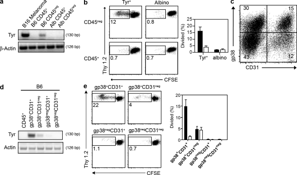
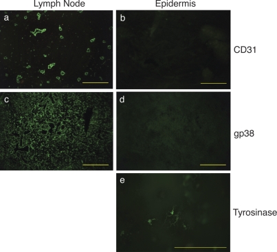
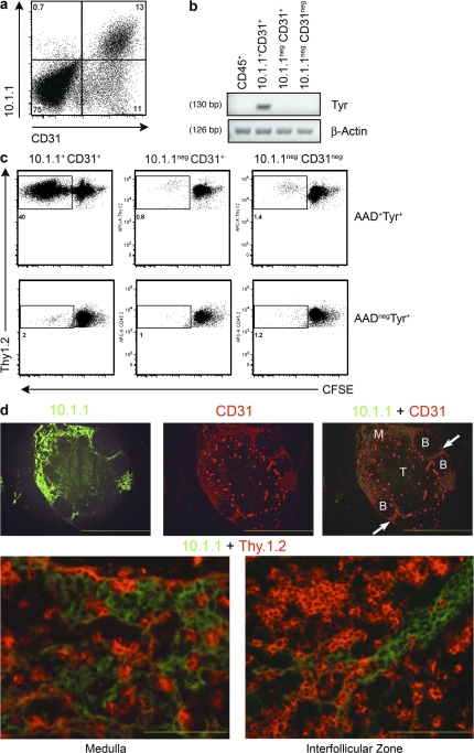
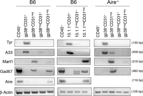
Peripheral immune tolerance is generally thought to result from cross-presentation of tissue-derived proteins by quiescent tissue-resident dendritic cells to self-reactive T cells that have escaped thymic negative selection, leading to anergy or deletion. Recently, we and others have implicated the lymph node (LN) stroma in mediating CD8 T cell peripheral tolerance. We demonstrate that LN-resident lymphatic endothelial cells express multiple peripheral tissue antigens (PTAs) independent of the autoimmune regulator (Aire). They directly present an epitope derived from one of these, the melanocyte-specific protein tyrosinase, to tyrosinase-specific CD8 T cells, leading to their deletion. We also show that other LN stromal subpopulations express distinct PTAs by mechanisms that vary in their Aire dependence. These results establish lymphatic endothelial cells, and potentially other LN-resident cells, as systemic mediators of peripheral immune tolerance.


Figures

Figure 1.
Figure 1.
gp38+CD31+ LN stromal cells express and present Tyr369 in an Aire-independent manner. (a) Tyrosinase (Tyr) and β-actin expression in CD45neg and CD45+ LN populations from B6 and albino animals was determined using 40-cycle RT-PCR. Data shown are representative of four independent experiments. (b) Proliferation of CFSE-labeled naive Thy1.2+ FH T cells co-cultured with Thy1.1+ CD45neg (black bars) or CD45+ (white bars) cells purified from the LNs of tyrosinase+ and albino animals was assessed after 86 h. Left, representative experiment; right, summary data for two independent experiments. (c) LN stromal cell populations defined based on expression of gp38 and CD31. The representative plot is gated on CD45neg cells. Numbers indicate percentage of total CD45neg cells. (d) Tyrosinase and β-actin expression in CD45+ cells and four LN stromal cell subpopulations identified by differential expression of gp38 and CD31. Purified populations were obtained by electronic cell sorting and 40-cycle RT-PCR was performed. Data shown are representative of six independent experiments. (e) Proliferation of CFSE-labeled naive Thy1.2+ FH T cells co-cultured with Thy1.1+ cells of the indicated purified LN stromal cell subpopulations from tyrosinase+ (black bars) and albino (white bars) animals was assessed after 86 h. Left, representative experiment; right, summary data for three independent experiments. Error bars indicate SEM.
Figure 2.
Figure 2.
The gp38+CD31+ LN stromal cells responsible for mediating tolerance to Tyr369 are not related to melanocytes. Immunofluorescence of a normal LN section (a and c) and epidermal ear sheets (b, d, and e) stained for the expression of LN stromal cell markers CD31 (100×; a and b) and gp38 (100×; c and d). Melanocytes in epidermal ear sheets were visualized by their tyrosinase staining (200×; e). Staining is representative of multiple magnifications and fields from two independent experiments consisting of two separate LNs and ears from three mice. Bars, 200 µm.
Figure 3.
Figure 3.
The LN stromal cells responsible for mediating tolerance to Tyr369 have characteristics of lymphatic endothelial cells. (a) Staining of CD45neg LN stromal cells with antibodies against 10.1.1 and CD31. (b) RT-PCR for tyrosinase (Tyr) and β-actin mRNA in CD45+ cells and the indicated LN stromal cell subpopulations was evaluated as described in Fig. 1. Results shown are representative of five independent experiments. (c) Proliferation of CFSE-labeled naive Thy1.2+ FH T cells co-cultured with cells of the indicated LN stromal cell subpopulations from Thy1.1+ tyrosinase+ and albino animals was assessed as in Fig. 1. Results are from two independent experiments performed with stromal cells isolated from six pooled mice each. (d, Top) Cells with dual staining of 10.1.1 and CD31 are located in the medulla (M) and in the interfollicular areas (arrows) but not in the T cell zone (T) or B cell follicles (B; 40×). Staining is representative of multiple magnifications and fields from two independent experiments consisting of two separate LNs from two mice. Bars, 1 mm. (d, Bottom) Higher magnification (400×) of the medulla (left) and the interfollicular region (right) showing intermingling of 10.1.1+ LEC and Thy1.1+ T cells. Staining is representative of multiple magnifications and fields from two independent experiments consisting of two separate LNs from one mouse. Bars, 400 µm.
Figure 4.
Figure 4.
LN stromal cell subsets express distinct groups of PTA that vary in their dependence on Aire. Tyrosinase (Tyr), A33, Mart1, Gad67, Aire, and β-actin mRNAs were amplified by 40-cycle RT-PCR from the indicated subpopulations of LN stromal cells isolated from either C57BL/6 or Aire−/− mice. Data from C57BL/6 mice sorted with gp38 and CD31 are representative of six independent experiments. Data from C57BL/6 mice sorted with 10.1.1 and CD31 are representative of three independent experiments. Data from Aire−/− mice sorted with gp38 and CD31 are representative of two independent experiments.

Comment in

References

    1. Belz G.T., Behrens G.M., Smith C.M., Miller J.F., Jones C., Lejon K., Fathman C.G., Mueller S.N., Shortman K., Carbone F.R., Heath W.R. 2002. The CD8α+ dendritic cell is responsible for inducing peripheral self-tolerance to tissue-associated antigens. J. Exp. Med. 196:1099–1104 10.1084/jem.20020861 - DOI - PMC - PubMed
    1. Colella T.A., Bullock T.N.J., Russell L.B., Mullins D.W., Overwijk W.W., Luckey C.J., Pierce R.A., Restifo N.P., Engelhard V.H. 2000. Self-tolerance to the murine homologue of a tyrosinase-derived melanoma antigen: implications for tumor immunotherapy. J. Exp. Med. 191:1221–1232 10.1084/jem.191.7.1221 - DOI - PMC - PubMed
    1. Derbinski J., Schulte A., Kyewski B., Klein L. 2001. Promiscuous gene expression in medullary thymic epithelial cells mirrors the peripheral self. Nat. Immunol. 2:1032–1039 10.1038/ni723 - DOI - PubMed
    1. Derbinski J., Gäbler J., Brors B., Tierling S., Jonnakuty S., Hergenhahn M., Peltonen L., Walter J., Kyewski B. 2005. Promiscuous gene expression in thymic epithelial cells is regulated at multiple levels. J. Exp. Med. 202:33–45 10.1084/jem.20050471 - DOI - PMC - PubMed
    1. DeVoss J., Hou Y., Johannes K., Lu W., Liou G.I., Rinn J., Chang H., Caspi R.R., Caspi R., Fong L., Anderson M.S. 2006. Spontaneous autoimmunity prevented by thymic expression of a single self-antigen. J. Exp. Med. 203:2727–2735 10.1084/jem.20061864 - DOI - PMC - PubMed

Publication types

MeSH terms