. 2010 Mar 22;188(6):833-49.
doi: 10.1083/jcb.200909117.

The TWEAK-Fn14 system is a critical regulator of denervation-induced skeletal muscle atrophy in mice

Affiliations

The TWEAK-Fn14 system is a critical regulator of denervation-induced skeletal muscle atrophy in mice

Ashwani Mittal et al. J Cell Biol. .

Abstract

Skeletal muscle atrophy occurs in a variety of clinical settings, including cachexia, disuse, and denervation. Inflammatory cytokines have been shown to be mediators of cancer cachexia; however, the role of cytokines in denervation- and immobilization-induced skeletal muscle loss remains unknown. In this study, we demonstrate that a single cytokine, TNF-like weak inducer of apoptosis (TWEAK), mediates skeletal muscle atrophy that occurs under denervation conditions. Transgenic expression of TWEAK induces atrophy, fibrosis, fiber-type switching, and the degradation of muscle proteins. Importantly, genetic ablation of TWEAK decreases the loss of muscle proteins and spared fiber cross-sectional area, muscle mass, and strength after denervation. Expression of the TWEAK receptor Fn14 (fibroblast growth factor-inducible receptor 14) and not the cytokine is significantly increased in muscle upon denervation, demonstrating an unexpected inside-out signaling pathway; the receptor up-regulation allows for TWEAK activation of nuclear factor kappaB, causing an increase in the expression of the E3 ubiquitin ligase MuRF1. This study reveals a novel mediator of skeletal muscle atrophy and indicates that the TWEAK-Fn14 system is an important target for preventing skeletal muscle wasting.


Figures

Figure 1.
Figure 1.
Characterization of TWEAK-Tg and TWEAK-KO mice. (A) Fold change in mRNA levels of TWEAK in skeletal muscle and other organs of TWEAK-Tg mice (n = 6) compared with littermate control (n = 6) mice. (B) Fold change in protein levels of TWEAK in skeletal muscle, other organs, and serum of TWEAK-Tg (n = 3) mice compared with control (n = 3) mice. (A and B) *, P < 0.01 (values significantly different from littermate control mice). (C) Fold change in the mRNA levels of TWEAK receptor Fn14 in TA muscle of TWEAK-Tg (n = 5) and TWEAK-KO (n = 6) mice compared with control (n = 6) mice. *, P < 0.01 (values significantly different from age-matched wild-type mice). (D) Fold change in mRNA levels of TNF, IL-1β, and IL-6 in TA muscle of TWEAK-Tg (n = 3) mice compared with control mice (n = 3). (E) Serum CK levels in TWEAK-Tg or TWEAK-KO mice compared with control mice measured using a CK assay kit (Stanbio Laboratory). (A–E) Error bars represent the SD. Dashed lines represent the basal levels in control mice.
Figure 2.
Figure 2.
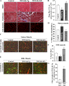
Effect of TWEAK on skeletal muscle phenotypes in vivo. (A) Soleus muscle from 6-mo-old control (n = 6), TWEAK-Tg (n = 8), and TWEAK-KO (n = 5) mice was analyzed after H&E or Masson’s trichrome staining or after immunostaining with laminin antibody. (B) Quantification of mean fiber CSA in soleus muscle of control, TWEAK-Tg, and TWEAK-KO mice. *, P < 0.01; and #, P < 0.05 (value significantly different from control mice). (C) Representative photomicrographs of soleus muscle sections from 6-mo-old control (n = 6), TWEAK-Tg (n = 7), and TWEAK-KO (n = 6) mice taken after immunostaining with anti–MyHC fast type and antilaminin. (D) Quantification of MyHC fast type–positive (filled with green color in C) and -negative fibers (empty) in control, TWEAK-Tg, and TWEAK-KO mice. (E) Representative photomicrographs of EDL muscle sections from 6-mo-old control (n = 4), TWEAK-Tg (n = 4), and TWEAK-KO (n = 4) mice taken after immunostaining with anti–type I and antilaminin. (F) Percentage of type I fibers (filled with red color in E) in control, TWEAK-Tg, and TWEAK-KO mice. (D and F) */#, P < 0.05 (values significantly different from control mice). (G) Measurement of mean fiber CSA of fast- and slow-type fibers in control (n = 5) and TWEAK-Tg (n = 5) mice. *, P < 0.01 (value significantly different from slow-type fibers). (B, D, F, and G) Error bars represent the SD. Bars, 50 µm.
Figure 3.
Figure 3.
Effect of TWEAK on the expression of specific muscle proteins in vivo. (A) Representative Western blots for MyHC, nNOS, tropomyosin, troponin, sarcomeric α-actin, dystrophin, laminin, and tubulin in soleus muscle of 6-mo-old control and TWEAK-Tg mice. (B) Representative Western blots for various muscle proteins in soleus muscle of 6-mo-old control and TWEAK-KO mice. (A and B) Black lines indicate that intervening lanes have been spliced out. (C) Fold change in protein levels of MyHC and nNOS in soleus muscle of control (n = 6), TWEAK-Tg (n = 5), and TWEAK-KO (n = 6) mice. */#, P < 0.05 (values significantly different from control mice). (D) Fold change in mRNA levels of MyHC and nNOS in soleus muscle of control (n = 6), TWEAK-Tg (n = 5), and TWEAK-KO (n = 4) mice. (E) Activation of NF-κB but not AP-1 transcription factor in soleus muscle of 6-mo-old TWEAK-Tg compared with littermate control mice. (F) Fold difference in mRNA levels of MAFbx and MuRF1 in control and TWEAK-Tg mice. *, P < 0.01 (values significantly different from level of MuRF1 in control mice). (C, D, and F) Error bars represent the SD. Dashed lines represent the basal levels in control mice.
Figure 4.
Figure 4.
Expression of TWEAK and Fn14 in skeletal muscle upon denervation. (A) Expression of Fn14 in skeletal muscle in response to casting (Cast.), denervation (Den.), and dexamethasone (Dex.) treatment to induce atrophy and in response to clenbuterol (Clen.) and recovery (Recov.) from casting to induce hypertrophy. Animals were also studied under free-running exercise conditions (exercise). mRNA was taken at the time points indicated and assessed in an Affymetrix microarray study. mRNA from 10 animals (n = 10) were used for each condition. (B) Relative mRNA levels of TWEAK, Fn14, and TNFR1 and -2 in denervated GA muscle versus sham-operated contralateral GA muscle after 4 d from 12-wk-old C57BL/6 mice (n = 5). *, P < 0.01. (A and B) Error bars represent the SD. Dashed lines represent the basal levels in control mice. (C) Levels of Fn14 protein measured by Western blotting in control (C) and denervated (D) GA muscle of C57BL/6 mice at 4, 7, or 10 d of denervation. (D) Expression of Fn14 protein in GA, TA, soleus, and EDL muscle of mice measured 4 d after denervation. The black line indicates that intervening lanes have been spliced out. (E) Representative Western blots showing expression of slow-type (st) MyHC (using clone A4.840) and fast-type (ft) MyHC (using clone BF-F3) in GA, TA, soleus, and EDL muscle of mice.
Figure 5.
Figure 5.
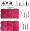
Role of TWEAK in denervation-induced skeletal muscle atrophy. (A) 3-mo-old control, TWEAK-Tg, and TWEAK-KO mice 10 d after denervation procedure. Arrows point to denervated GA muscle. (B) TA and soleus muscle were isolated from tendon to tendon from control, TWEAK-Tg, and TWEAK-KO mice 10 d after denervation (n = 6 per group), and their wet weight was measured. */#, P < 0.05 (values significantly different from corresponding muscle of control mice). (C) H&E-stained sections of TA of control, TWEAK-Tg, and TWEAK-KO mice 10 d after denervation. (D) Quantification of fiber CSA of TA muscle in control, TWEAK-Tg, and TWEAK-KO mice 10 d after denervation (n = 8 in each group). (E) Representative photomicrographs of H&E-stained soleus muscle sections from control, TWEAK-Tg, and TWEAK-KO mice 12 d after denervation. (F) Measurement of fiber CSA in H&E-stained soleus muscle sections in control, TWEAK-Tg, and TWEAK-KO mice 12 d after denervation (n = 6 in each group). (D and F) *, P < 0.01; and #, P < 0.05 (values significantly different from that of control mice at indicated time after denervation). (B, D, and F) Error bars represent the SD. Bars, 20 µm.
Figure 6.
Figure 6.
Role of TWEAK in development of fibrosis and loss of muscle function. (A) Sirius red staining performed on TA muscle sections after 21 d of denervation in control, TWEAK-Tg, and TWEAK-KO mice (n = 4 in each group). (B) Fold change in the mRNA levels of collagen type 1, α 2 (Col1a2) in TA and soleus muscle of control (n = 6), TWEAK-Tg (n = 3), and TWEAK-KO (n = 3) mice 10 d after denervation measured by QRT-PCR. */#, P < 0.01 (values significantly different from denervated muscle of control mice). (C) Denervation-induced loss in absolute muscle force production in isometric contraction was measured in soleus muscle of control (n = 5) and TWEAK-KO (n = 6) mice at 80, 120, 150, 220, and 300 Hz. *, P < 0.01 (values significantly different from the denervated soleus muscle of control mice at same frequency). (B and C) Error bars represent the SD. Bars, 50 µm.
Figure 7.
Figure 7.
TWEAK-neutralizing antibody rescues denervation-induced muscle loss in mice. 2-mo-old C57BL/6 mice were denervated for 2 d, followed by intraperitoneal injections of 200 µg/mouse of either rat IgG1 or anti-TWEAK every third day for 12 d (n = 4 in each group). (A) Representative photomicrographs of H&E-stained sections of control and denervated TA muscle of isotype and anti-TWEAK–treated mice. (B) Quantification of fiber CSA in TA muscle sections of isotype and anti-TWEAK–treated mice. *, P < 0.01 (values significantly different from denervated TA muscle of IgG1-treated mice). (C) H&E-stained sections of control and denervated soleus muscle of mice treated with isotype or anti-TWEAK. (D) Quantification of fiber CSA in soleus muscle sections. #, P < 0.05 (values significantly different from denervated soleus muscle of IgG1-treated mice). (B and D) Error bars represent the SD. ITCT, isotype control. Bars, 20 µm.
Figure 8.
Figure 8.
Role of TWEAK in the activation of NF-κB in denervated skeletal muscles. DNA-binding activity of NF-κB was measured by EMSA in TA muscle 10 d after denervation. (A and B) Representative EMSA gel from three independent experiments for control and TWEAK-Tg mice (A) and control and TWEAK-KO mice (B). (C) Supershift assay performed using nuclear extracts from denervated GA muscle of control mice using antibodies against p50 and p52 subunits of NF-κB. (D) Fold change in NF-κB reporter gene activity (normalized using Renilla luciferase) in TA muscle of control, TWEAK-Tg, and TWEAK-KO mice after denervation. *, P < 0.01; and #, P < 0.05 (values significantly different from corresponding control mice). Error bars represent the SD. U, undenervated; D, denervated; PIS, preimmune serum.
Figure 9.
Figure 9.
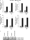
Effects of TWEAK on the expression of muscle-specific E3 ubiquitin ligases in denervated skeletal muscle. (A) Relative mRNA levels of MAFbx in denervated GA muscle of TWEAK-Tg or TWEAK-KO versus control mice (n = 6 in each group). */#, P < 0.01 (values significantly different from corresponding control mice). (B) Relative mRNA levels of MuRF1 in denervated GA muscle of TWEAK-Tg or TWEAK-KO versus control mice (n = 6 per group). (A and B) Error bars represent the SD. (C) Representative immunoblots demonstrating MuRF1 protein levels in undenervated (U) and denervated (D) GA muscle of control, TWEAK-Tg, and TWEAK-KO mice. Black lines indicate that intervening lanes have been spliced out.

References

    1. Acharyya S., Guttridge D.C. 2007. Cancer cachexia signaling pathways continue to emerge yet much still points to the proteasome. Clin. Cancer Res. 13:1356–1361 10.1158/1078-0432.CCR-06-2307 - DOI - PubMed
    1. Acharyya S., Ladner K.J., Nelsen L.L., Damrauer J., Reiser P.J., Swoap S., Guttridge D.C. 2004. Cancer cachexia is regulated by selective targeting of skeletal muscle gene products. J. Clin. Invest. 114:370–378 - PMC - PubMed
    1. Acharyya S., Butchbach M.E., Sahenk Z., Wang H., Saji M., Carathers M., Ringel M.D., Skipworth R.J., Fearon K.C., Hollingsworth M.A., et al. 2005. Dystrophin glycoprotein complex dysfunction: a regulatory link between muscular dystrophy and cancer cachexia. Cancer Cell. 8:421–432 10.1016/j.ccr.2005.10.004 - DOI - PubMed
    1. Argilés J.M., Busquets S., López-Soriano F.J. 2005. The pivotal role of cytokines in muscle wasting during cancer. Int. J. Biochem. Cell Biol. 37:2036–2046 10.1016/j.biocel.2005.03.014 - DOI - PubMed
    1. Attaix D., Bechet D. 2007. FoxO3 controls dangerous proteolytic liaisons. Cell Metab. 6:425–427 10.1016/j.cmet.2007.11.005 - DOI - PubMed

Publication types

MeSH terms