

Prostate cancer: value of multiparametric MR imaging at 3 T for detection--histopathologic correlation
- PMID: 20308447
- PMCID: PMC2843833
- DOI: 10.1148/radiol.09090475
Prostate cancer: value of multiparametric MR imaging at 3 T for detection--histopathologic correlation
Abstract
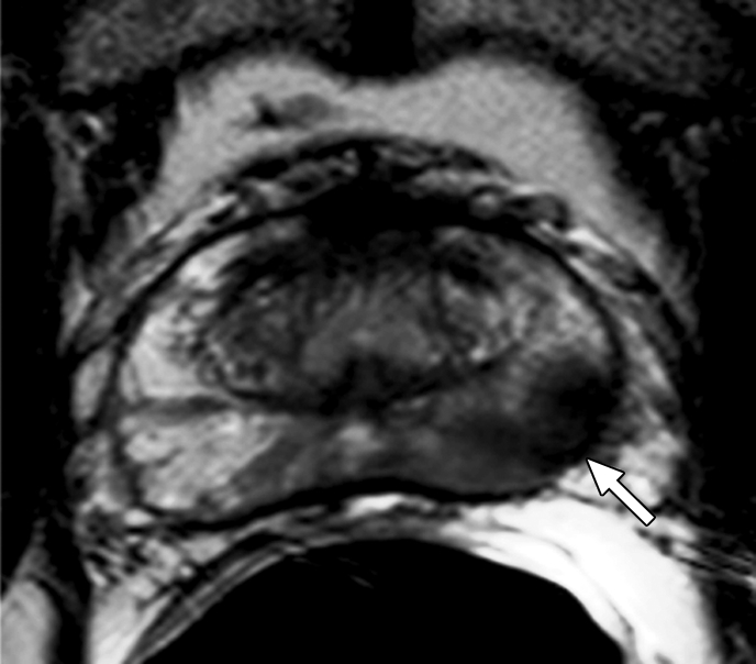
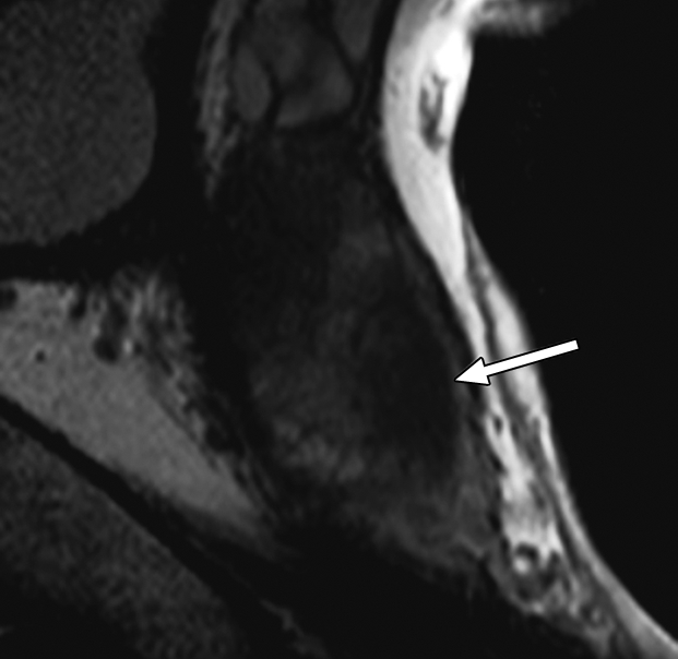
Purpose: To determine utility of multiparametric imaging performed at 3 T for detection of prostate cancer by using T2-weighted magnetic resonance (MR) imaging, MR spectroscopy, and dynamic contrast material-enhanced MR imaging, with whole-mount pathologic findings as reference standard.
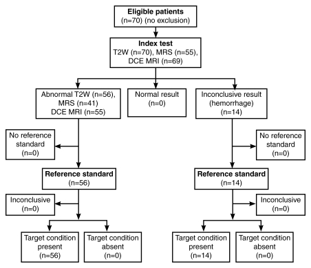
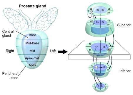
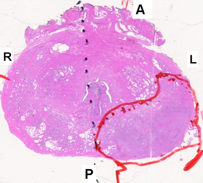
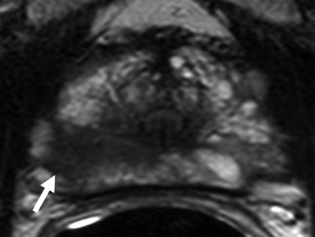
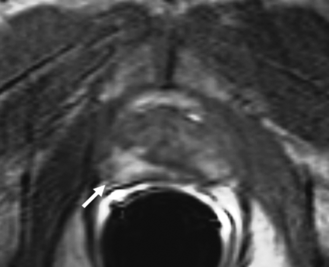
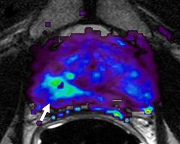
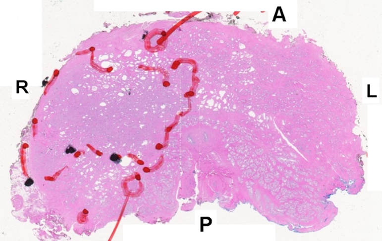
Materials and methods: This prospectively designed, HIPAA-compliant, single-institution study was approved by the local institutional review board. Seventy consecutive patients (mean age, 60.4 years; mean prostate-specific antigen level, 5.47 ng/mL [5.47 microg/L]; range, 1-19.9 ng/mL [1-19.9 microg/L]) were included; informed consent was obtained from each patient. All patients had biopsy-proved prostate cancer, with a median Gleason score of 7 (range, 6-9). Images were obtained by using a combination of six-channel cardiac and endorectal coils. MR imaging and pathologic findings were evaluated independently and blinded and then correlated with histopathologic findings by using side-by-side comparison. Analyses were conducted with a raw stringent approach and an alternative neighboring method, which accounted for surgical deformation, shrinkage, and nonuniform slicing factors in pathologic specimens. Generalized estimating equations (GEEs) were used to estimate the predictive value of region-specific, pathologically determined cancer for all three modalities. This approach accounts for the correlation among multiple regions in the same individual.
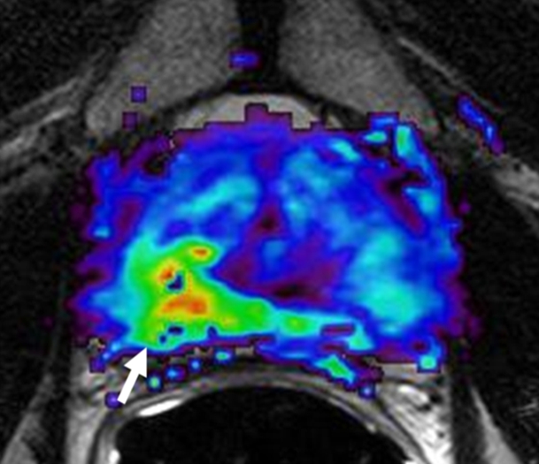
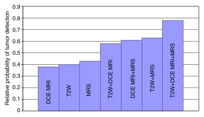
Results: For T2-weighted MR imaging, sensitivity and specificity values obtained with stringent approach were 0.42 (95% confidence interval [CI]: 0.36, 0.47) and 0.83 (95% CI: 0.81, 0.86), and for the alternative neighboring approach, sensitivity and specificity values were 0.73 (95% CI: 0.67, 0.78) and 0.89 (95% CI: 0.85, 0.93), respectively. The combined diagnostic accuracy of T2-weighted MR imaging, dynamic contrast-enhanced MR imaging, and MR spectroscopy for peripheral zone tumors was examined by calculating their predictive value with different combinations of techniques; T2-weighted MR imaging, dynamic contrast-enhanced MR imaging, and MR spectroscopy provided significant independent and additive predictive value when GEEs were used (P < .001, P = .02, P = .002, respectively).
Conclusion: Multiparametric MR imaging (T2-weighted MR imaging, MR spectroscopy, dynamic contrast-enhanced MR imaging) of the prostate at 3 T enables tumor detection, with reasonable sensitivity and specificity values.
RSNA, 2010
Figures
References
-
- American Cancer Society Cancer facts & figures 2009 Atlanta, Ga: American Cancer Society, 2009
-
- Kim CK, Park BK, Kim B. Localization of prostate cancer using 3T MRI: comparison of T2-weighted and dynamic contrast-enhanced imaging. J Comput Assist Tomogr 2006;30(1):7–11 - PubMed
-
- Torricelli P, Cinquantini F, Ligabue G, Bianchi G, Sighinolfi P, Romagnoli R. Comparative evaluation between external phased array coil at 3 T and endorectal coil at 1.5 T: preliminary results. J Comput Assist Tomogr 2006;30(3):355–361 - PubMed
-
- Park BK, Kim B, Kim CK, Lee HM, Kwon GY. Comparison of phased-array 3.0-T and endorectal 1.5-T magnetic resonance imaging in the evaluation of local staging accuracy for prostate cancer. J Comput Assist Tomogr 2007;31(4):534–538 - PubMed
-
- Fütterer JJ, Heijmink SW, Scheenen TW, et al. Prostate cancer localization with dynamic contrast-enhanced MR imaging and proton MR spectroscopic imaging. Radiology 2006;241(2):449–458 - PubMed
Publication types
MeSH terms
Substances
Grants and funding
LinkOut - more resources
Full Text Sources
Other Literature Sources
Medical
