

Head and neck cancers on CT: preliminary study of treatment response assessment based on computerized volume analysis
- PMID: 20308515
- PMCID: PMC3729396
- DOI: 10.2214/AJR.09.2817
Head and neck cancers on CT: preliminary study of treatment response assessment based on computerized volume analysis
Abstract
Objective: The objective of our study was to investigate the feasibility of computerized segmentation of lesions on head and neck CT scans and evaluate its potential for estimating changes in tumor volume in response to treatment of head and neck cancers.
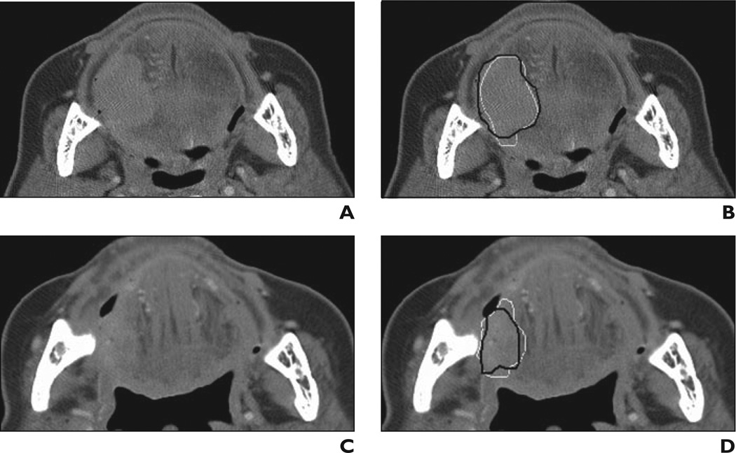
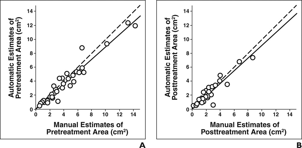
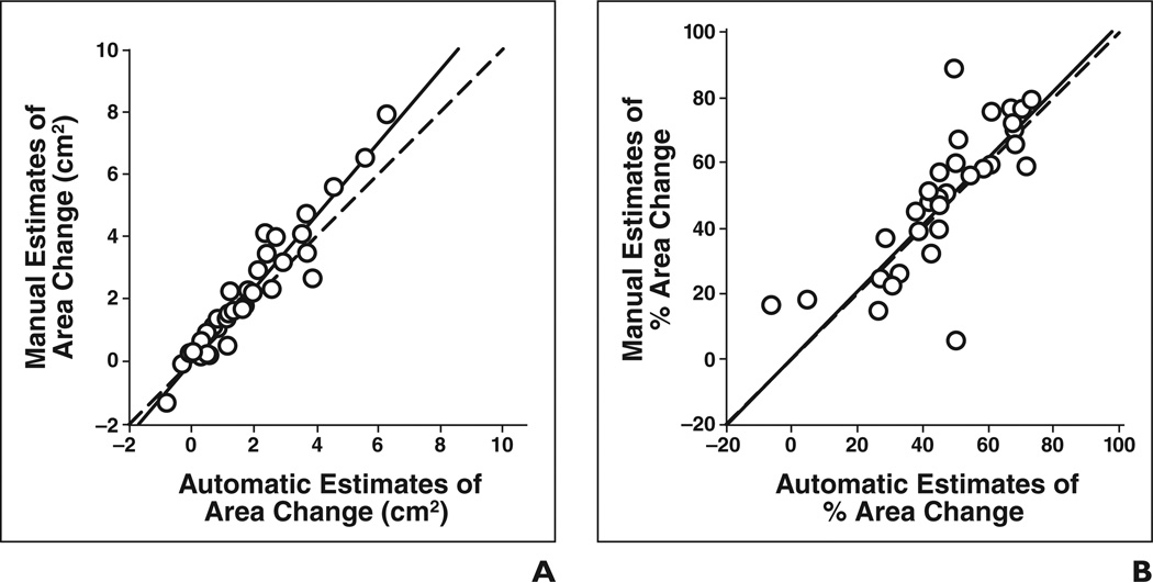
Materials and methods: Twenty-six CT scans were retrospectively collected from the files of 13 patients with 35 head and neck lesions. The CT scans were obtained from an examination performed before treatment (pretreatment scan) and an examination performed after one cycle of chemotherapy (posttreatment scan). Thirteen lesions were primary site cancers and 22 were metastatic lymph nodes. An experienced radiologist (radiologist 1) marked the 35 lesions and outlined each lesion's 2D contour on the best slice on both the pre- and posttreatment scans. Full 3D contours were also manually extracted for the 13 primary tumors. Another experienced radiologist (radiologist 2) verified and modified, if necessary, all manually drawn 2D and 3D contours. An in-house-developed computerized system performed 3D segmentation based on a level set model.
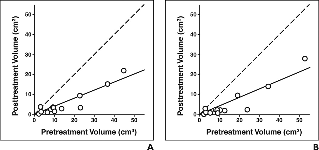
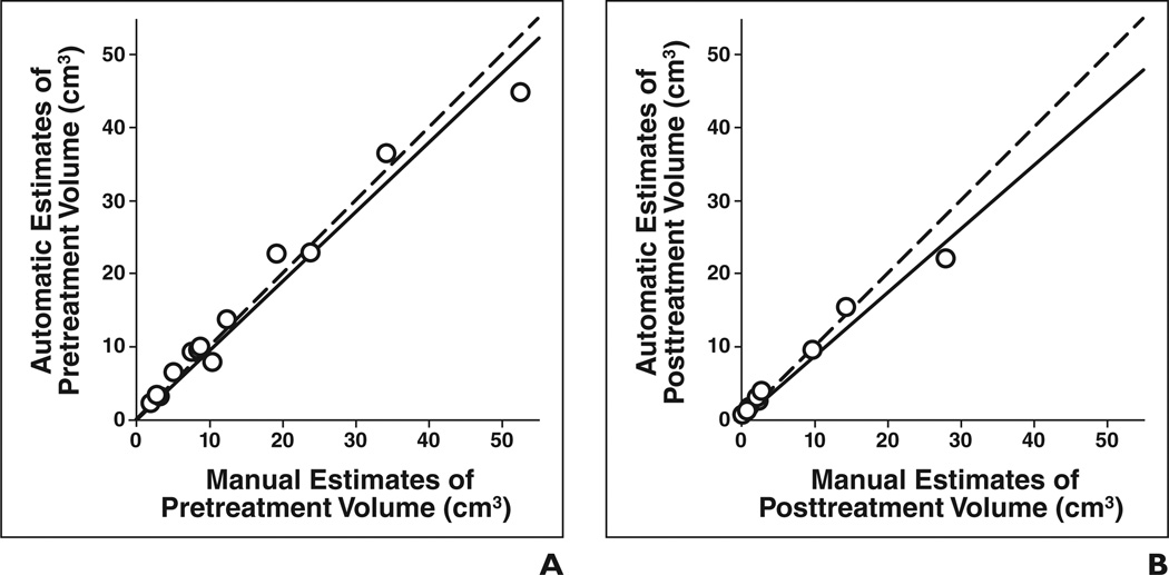
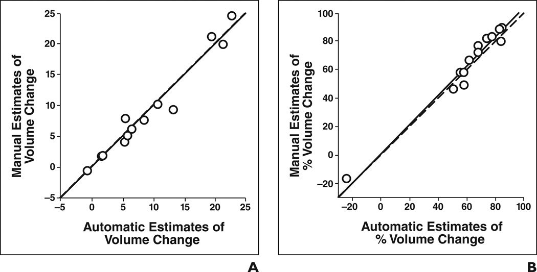
Results: The computer-estimated change in tumor volume and percentage change in tumor volume between the pre- and posttreatment scans achieved a high correlation (intraclass correlation coefficient [ICC] = 0.98 and 0.98, respectively) with the estimates from manual segmentation for the 13 primary tumors. The average error in estimating the percentage change in tumor volume by automatic segmentation relative to the radiologists' average error was -1.5% +/- 5.4% (SD). For the 35 lesions, the ICC between the automatic and manual estimates of change in pre- to posttreatment tumor area was 0.93 and of percentage change in pre- to posttreatment tumor area was 0.85. The average error in estimating the percentage change in tumor area by automatic segmentation was -3.2% +/- 15.3%.
Conclusion: Preliminary results indicate that this computerized segmentation system can reliably estimate changes in tumor size on CT scans relative to radiologists' manual segmentation. This information can be used to calculate changes in tumor size on pre- and posttreatment scans to assess response to treatment.
Figures
References
-
- American Cancer Society. Cancer facts & figures 2007. Atlanta, GA: American Cancer Society; 2007.
-
- Induction chemotherapy plus radiation compared with surgery plus radiation in patients with advanced laryngeal cancer. The Department of Veterans Affairs Laryngeal Cancer Study Group. N Engl J Med. 1991;324:1685–1690. [No authors listed]. - PubMed
-
- Lefebvre JL, Chevalier D, Luboinski B, Kirkpatrick A, Collette L, Sahmoud T. Larynx preservation in pyriform sinus cancer: preliminary results of a European organization for research and treatment of cancer phase III trial. J Natl Cancer Inst. 1996;88:890–899. - PubMed
-
- Mancuso AA, Mukherji SK, Schmalfuss I, et al. Preradiotherapy computed tomography as a predictor of local control in supraglottic carcinoma. J Clin Oncol. 1999;17:631–637. - PubMed
-
- Chua DT, Sharn JS, Kwong DL, et al. Volumetric analysis of tumor extent in nasopharyngeal carcinoma and correlation with treatment outcome. Int J Radiat Oncol Biol Phys. 1997;39:711–719. - PubMed
Publication types
MeSH terms
Substances
Grants and funding
LinkOut - more resources
Full Text Sources
Medical
