. 2010 Jun;151(6):2923-32.
doi: 10.1210/en.2010-0081. Epub 2010 Mar 22.

The nuclear receptor cofactor receptor-interacting protein 140 is a positive regulator of amphiregulin expression and cumulus cell-oocyte complex expansion in the mouse ovary

Affiliations

The nuclear receptor cofactor receptor-interacting protein 140 is a positive regulator of amphiregulin expression and cumulus cell-oocyte complex expansion in the mouse ovary

Jaya Nautiyal et al. Endocrinology. 2010 Jun.

Abstract

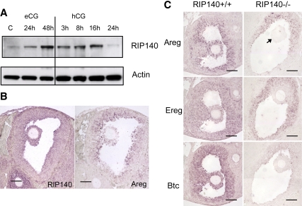
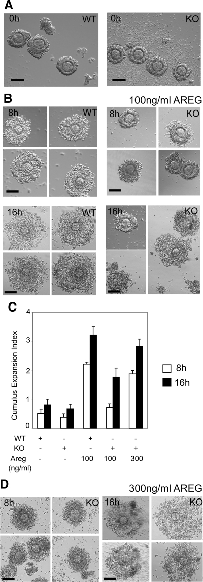
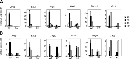
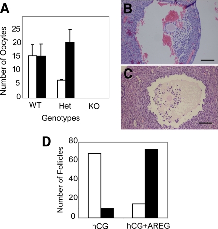
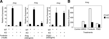
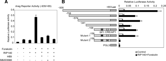
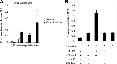
The nuclear receptor cofactor receptor-interacting protein 140 (RIP140) is essential for cumulus cell-oocyte complex (COC) expansion, follicular rupture, and oocyte release during ovulation. The expression of many genes necessary for COC expansion is impaired in the absence of RIP140, but the studies herein document that their expression can be restored and COC expansion rescued by treatment with the epidermal growth factor (EGF)-like factor amphiregulin (AREG) both in vitro and in vivo. We demonstrate by several approaches that RIP140 is required for the expression of the EGF-like factors in granulosa cells, but the dependence of genes involved in cumulus expansion, including Ptgs2 Has2, Tnfaip6, and Ptx3, is indirect because they are induced by AREG. Treatment of granulosa cells with forskolin to mimic the effects of LH increases AREG promoter activity in a RIP140-dependent manner that 1) requires an intact cAMP response element in the proximal promoter region of the Areg gene and 2) involves its actions as a coactivator for cAMP response element-binding protein/c-Jun transcription factors. Although human chorionic gonadotropin and AREG coadministration is sufficient to restore ovulation fully in RIP140 heterozygous mice in vivo, both follicular rupture and ovulation remain impaired in the RIP140 null mice. Thus, we conclude that although the level of RIP140 expression in the ovary is a crucial factor required for the transient expression of EGF-like factors necessary for cumulus expansion, it also plays a role in other signaling pathways that induce follicular rupture.


Figures

Figure 1
Figure 1
Expression of RIP140 and EGF-like factors in the ovary. A, Western blot analysis showing RIP140 protein from extracts obtained from ovaries of 3-wk-old mice, untreated or treated with eCG for 24 and 48 h and eCG for 48 h followed by hCG for 3, 8, 16, and 24 h (n = 2 for each treatment group). β-Actin was used as a loading control. B, In situ hybridization analysis showing expression of both RIP140 and Areg in mural and cumulus granulosa cells in ovary sections obtained from mice treated with eCG for 48 h followed by hCG for 3 h. C, In situ hybridization analysis to detect expression of Areg, Ereg, and Btc in WT and RIP140 knockout (KO) ovary sections obtained from mice treated with eCG for 48 h followed by hCG for 3 h. The RIP140 KO sections show a reduced expression of all three EGF-like factors in the granulosa cells and no expression in the cumulus cells (arrow). Scale bars, 100 μm. Images are representative of experiments done using three animals in each group.
Figure 2
Figure 2
Restoration of cumulus expansion in RIP140 null COCs. A, COCs released from WT and RIP140 knockout (KO) ovaries after 48-h eCG treatment. Scale bar, 100 μm. B, COCs isolated from WT and RIP140 KO ovaries after 48-h eCG treatment and cultured for 8 and 16 h in the presence of 100 ng/ml AREG. Scale bar, 100 μm. Compared with the WT COCs, the KO COCs showed defects in expansion at 8 h (P < 0.0001) with some COCs showing matrix formation by 16 h. C, Cumulus expansion index scored for COCs. D, COCs isolated from RIP140 KO ovaries after 48-h eCG treatment and cultured for 8 and 16 h in the presence of 300 ng/ml AREG showed improvement in cumulus expansion at 8 h and 16 h. Scale bar, 100 μm. Each treatment was performed with a pool of eight to ten COCs isolated from two to three mice, and three to four independent experiments were performed.
Figure 3
Figure 3
Time-course expression profile of genes involved in cumulus expansion. COCs were isolated from WT and RIP140 knockout (KO) mice primed with eCG for 48 h and cultured for 4, 8, and 16 h with either 100 ng/ml AREG (A) or 300 ng/ml AREG (B). RNA was isolated from the cultured COCs, and the expression of genes assessed using Q-PCR. Expression of all analyzed genes was induced in WT COCs by 100 ng/ml AREG in a time course. In contrast, 100 ng/ml AREG failed to induce the expression of these genes in the KO COCs. The expression of Ptgs2, Has2, Tnfαip6, and Ptx3 was remarkably induced in the KO COCs cultured with 300 ng/ml AREG with impaired induction of Areg and Ereg. Most treatment groups represent up to four independent experiments containing pools of eight to ten COCs.
Figure 4
Figure 4
Effect of AREG on ovulation in vivo. Coadministration of AREG and hCG in vivo rescues ovulation from RIP140 heterozygous mice but not RIP140 null mice. A, COCs recovered from the oviduct were counted after treating WT, RIP140 heterozygous, and RIP140 null mice with hCG for 16 h (open bars) or hCG plus 300 ng/ml AREG (closed bars) for 16 h (n = 3–4 for each group). Heterozygous animals showed an increase in ovulation on administration of AREG (P < 0.04). To analyze in vivo cumulus expansion, serial ovarian sections were analyzed from RIP140 null mice treated with hCG for 16 h (B) or hCG plus 300 ng/ml AREG for 16 h (C). Scale bar, 100 μm. D, Number of unexpanded (open bars) and expanded (closed bars) COCs counted from RIP140 null ovaries treated with hCG or hCG plus 300 ng/ml AREG for 16 h (n = 3 for each group).
Figure 5
Figure 5
Regulation of Areg in primary granulosa cells. WT and RIP140 null mice were treated with eCG for 48 h and granulosa cells (A) and COCs (B), isolated from large follicles, and cultured for 4 and 8 h, respectively, in the absence or presence of 10 μm forskolin, 300 ng/ml AREG, or 500 ng/ml PGE2. Areg expression relative to the untreated controls was determined by real-time PCR (n = 3 for each treatment; B represents data obtained from COCs pooled from three different animals). Areg expression in the RIP140 knockout (KO) granulosa cells and COCs was reduced in the presence of forskolin and AREG.
Figure 6
Figure 6
RIP140 acts as a coactivator of Areg. A, Reporter activity of a −935/+65 fragment of the Areg promoter fused to a luciferase reporter, transiently transfected in WTF4 granulosa cells together with either pCIEF-RIP140 or pCIEF empty vector. After 24-h transfection, cells were treated in the presence or absence of forskolin with or without PKA inhibitor, H89 (10 μm), or p38 MAPK inhibitor SB203580 (20 μm) for 4 h. In the inhibitor-treated groups, the inhibitors were added 30 min before forskolin stimulation (n = 8). B, Reporter activity of a series of Areg deletion constructs and point mutants fused to a luciferase reporter, transiently transfected in WTF4 granulosa cells with either pCIEF or pCIEF-RIP140 in the presence of forskolin (10 μm). pGL3 basic was used as a control. Schematic representations of the WT and mutated promoter constructs are shown.
Figure 7
Figure 7
RIP140 is directly recruited to the Areg promoter together with CREB and c-Jun. A, ChIP assays were performed in WTF4 granulosa cells in unstimulated and cells stimulated with forskolin (10 μm), using antibodies specific for RIP140, p-CREB, and c-Jun or nonspecific IgG. Precipitated fragments were analyzed by real-time PCR using Areg primers in the proximity of the CRE (n = 2). B, WTF4 granulosa cells were transiently transfected with siRNA oligos for c-Jun, CREB, or control, and after 48 h, cells were cotransfected with the Areg reporter gene (−935/+65), with either pCEIF or pCIEF-RIP140. After 24 h, cells were stimulated with forskolin for 4 h, and reporter activity was determined (n = 6).

Similar articles

Cited by

References

    1. Richards JS 2005 Ovulation: new factors that prepare the oocyte for fertilization. Mol Cell Endocrinol 234:75–79 - PubMed
    1. Richards JS 2007 Genetics of ovulation. Semin Reprod Med 25:235–242 - PubMed
    1. Russell DL, Robker RL 2007 Molecular mechanisms of ovulation: co-ordination through the cumulus complex. Hum Reprod Update 13:289–312 - PubMed
    1. Edson MA, Nagaraja AK, Matzuk MM 2009 The mammalian ovary from genesis to revelation. Endocr Rev 30:624–712 - PMC - PubMed
    1. Park JY, Su YQ, Ariga M, Law E, Jin SL, Conti M 2004 EGF-like growth factors as mediators of LH action in the ovulatory follicle. Science 303:682–684 - PubMed

Publication types

MeSH terms