. 2010 May;151(5):2349-60.
doi: 10.1210/en.2009-1227. Epub 2010 Mar 22.

Studies in zebrafish reveal unusual cellular expression patterns of gonadotropin receptor messenger ribonucleic acids in the testis and unexpected functional differentiation of the gonadotropins

Affiliations

Studies in zebrafish reveal unusual cellular expression patterns of gonadotropin receptor messenger ribonucleic acids in the testis and unexpected functional differentiation of the gonadotropins

Angel García-López et al. Endocrinology. 2010 May.

Abstract

This study aimed to improve, using the zebrafish model, our understanding of the distinct roles of pituitary gonadotropins FSH and LH in regulating testis functions in teleost fish. We report, for the first time in a vertebrate species, that zebrafish Leydig cells as well as Sertoli cells express the mRNAs for both gonadotropin receptors (fshr and lhcgr). Although Leydig cell fshr expression has been reported in other piscine species and may be a common feature of teleost fish, Sertoli cell lhcgr expression has not been reported previously and might be related to the undifferentiated gonochoristic mode of gonadal sex differentiation in zebrafish. Both recombinant zebrafish (rzf) gonadotropins (i.e. rzfLH and rzfFSH) stimulated androgen release in vitro and in vivo, with rzfFSH being significantly more potent than rzfLH. Forskolin-induced adenylate cyclase activation mimicked, whereas the protein kinase A inhibitor H-89 significantly reduced, the gonadotropin-stimulated androgen release. Therefore, we conclude that both FSH receptor and LH/choriogonadotropin receptor signaling are predominantly mediated through the cAMP/protein kinase A pathway to promote steroid production. Despite this similarity, other downstream mechanisms seem to differ. For example, rzfFSH up-regulated the testicular mRNA levels of a number of steroidogenesis-related genes both in vitro and in vivo, whereas rzfLH or human chorionic gonadotropin did not. Although not fully understood at present, these differences could explain the capacity of FSH to support both steroidogenesis and spermatogenesis on a long-term basis, whereas LH-stimulated steroidogenesis might be a more acute process, possibly restricted to periods during which peak steroid levels are required.


Figures
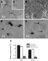
Figure 1
Figure 1
Cellular localization of fshr and lhcgr mRNA expression in zebrafish testis. A and C, In situ hybridization with either an antisense (A) or a sense (C) riboprobe for zebrafish fshr; note the positive staining in Leydig cells (Lc) in the interstitial tissue in A and the absence of signal in C. In both cases, germ cells were devoid of staining. B, Toluidine blue-stained 2-μm-thick plastic section showing groups of Leydig cells (Lc) within the interstitial compartment (enclosed by broken lines). D and E, Leydig cells are also identified by their positive staining with an insl3 antisense cRNA probe (D) and with the 3β-Hsd enzymatic reaction (E). Scale bars, 20 μm. F, Relative fshr and lhcgr mRNA expression levels in two microdissected testis tissue fractions (interstitial and intratubular) and a germ-cell-enriched population obtained from vas::EGFP transgenic zebrafish by FACS (germ cells only). Data correspond to values from two experiments, each with duplicate measurements (mean ± sem), normalized to β-actin1 mRNA levels, and expressed as percentage of fshr transcript amounts in the interstitial fraction. Note the logarithmic scale.
Figure 2
Figure 2
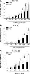
Stimulation of androgen release by zebrafish testicular explants. Amounts of 11-KT and OHA (mean ± sem) measured in incubation media after overnight (18 h) exposure to increasing concentrations of zfFSH (panel A), rzfLH (panel B), or the adenylate cyclase activator forskolin (panel C). B, Basal release. Values represent compiled data from two experiments, each with six replicates per ligand concentration. Different letters denote significant differences among groups (P < 0.05).
Figure 3
Figure 3
Effects of the PKA inhibitor H-89 on the gonadotropin-stimulated androgen release by zebrafish testicular explants. Amounts of 11-KT and OHA (mean ± sem) measured in incubation media after overnight (18 h) exposure to 250 ng/ml rzfFSH or 1000 ng/ml rzfLH alone and in combination with 100 μm H-89. Values represent compiled data from two experiments, each with six to seven replicates per condition. *, Values are significantly different (P < 0.05) from the respective gonadotropin-only condition.
Figure 4
Figure 4
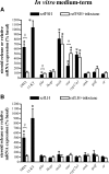
In vitro medium-term (2 d) modulation of androgen release and testicular gene expression by rzf gonadotropins. Amounts of the androgens OHA and 11-KT measured in incubation media and relative mRNA expression levels of several testicular genes (fshr, lhcgr, insl3, star, cyp17a1, amh, gsdf, and ar) after 2 d exposure to 100 ng/ml rzfFSH (A) or 500 ng/ml rzfLH (B) alone (black bars) and in combination with 25 μg/ml of the 3β-Hsd inhibitor trilostane (white bars). Data (mean ± sem) come from an experiment with eight replicates per condition and are expressed as percentage of basal levels, which were set to 100% for each parameter analyzed. Basal androgen release was 237 ± 18 pg OHA/mg tissue and 147 ± 17 pg 11-KT/mg tissue. Gene expression levels were normalized to β-actin1 mRNA levels. *, Values are significantly different (P < 0.05) from the respective basal condition in the absence of recombinant gonadotropin; #, significant difference (P < 0.05) between the absence and the presence of trilostane.
Figure 5
Figure 5
In vitro short-term (2 h) modulation of androgen release and testicular gene expression by rzf gonadotropins. Amounts of androgen (11-KT) measured in incubation media and relative mRNA expression levels of several testicular genes (fshr, lhcgr, insl3, star, cyp17a1, amh, gsdf, and ar) after 2 h exposure to 100 ng/ml rzfFSH (black bars) or 500 ng/ml rzfLH (white bars). Data (mean ± sem) come from an experiment with eight replicates per condition and are expressed as percentage of basal levels, which were set to 100% for each parameter analyzed. Basal androgen release was 40.0 ± 4.9 pg 11-KT/mg tissue. Gene expression levels were normalized to β-actin1 mRNA levels. *, Values are significantly different (P < 0.05) from the respective basal condition in the absence of recombinant gonadotropin; #, significant difference (P < 0.05) between treatment with rzfFSH and rzfLH.
Figure 6
Figure 6
In vivo short-term (2 h) modulation of plasma androgen levels and testicular gene expression by rzf gonadotropins and hCG. Circulating 11-KT concentrations and relative mRNA expression levels of several testicular genes (fshr, lhcgr, insl3, star, cyp17a1, amh, gsdf, and ar) 2 h after injection with 100 ng/g body weight rzfFSH (black bars), rzfLH (white bars) or 10 IU/g body weight hCG (gray bars). Data (mean ± sem) come from an experiment with eight fish per condition and are expressed as percentage of control levels, which were set to 100% for each parameter analyzed. Control androgen levels were 3.6 ± 0.9 ng 11-KT/ml plasma. Gene expression levels were normalized to β-actin1 mRNA levels. *, Values are significantly different (P < 0.05) from the basal control condition. Different letters denote significant differences among groups (P < 0.05).

Similar articles

Cited by

References

    1. Pierce JG, Parsons TF 1981 Glycoprotein hormones: structure and function. Annu Rev Biochem 50:465–495 - PubMed
    1. McLachlan RI, Wreford NG, O'Donnell L, de Kretser DM, Robertson DM 1996 The endocrine regulation of spermatogenesis: independent roles for testosterone and FSH. J Endocrinol 148:1–9 - PubMed
    1. Kumar TR 2005 What have we learned about gonadotropin function from gonadotropin subunit and receptor knockout mice? Reproduction 130:293–302 - PubMed
    1. Petersen C, Soder O 2006 The Sertoli cell: a hormonal target and ‘super’ nurse for germ cells that determines testicular size. Horm Res 66:153–161 - PubMed
    1. Saez JM 1994 Leydig cells: endocrine, paracrine, and autocrine regulation. Endocr Rev 15:574–626 - PubMed

Publication types

MeSH terms