. 2010 Apr 20;107(16):7509-14.
doi: 10.1073/pnas.0913199107. Epub 2010 Mar 22.

Short N-terminal sequences package proteins into bacterial microcompartments

Affiliations

Short N-terminal sequences package proteins into bacterial microcompartments

Chenguang Fan et al. Proc Natl Acad Sci U S A. .

Abstract

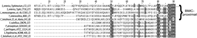
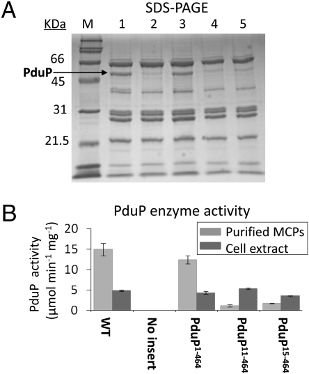
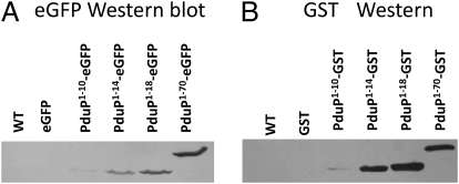
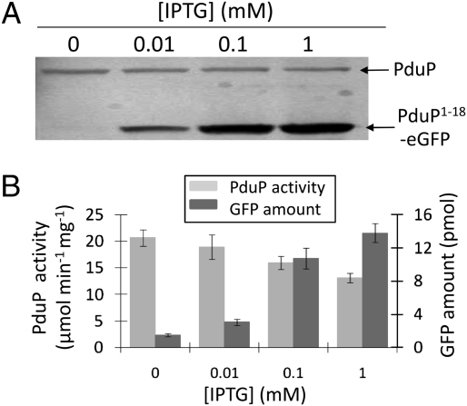
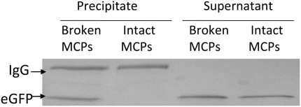
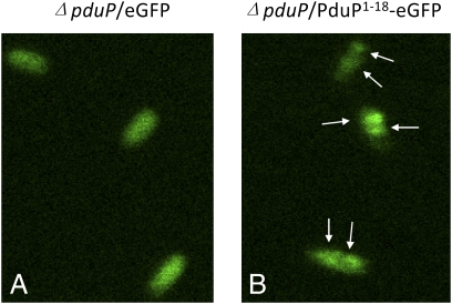
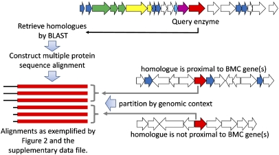
Hundreds of bacterial species produce proteinaceous microcompartments (MCPs) that act as simple organelles by confining the enzymes of metabolic pathways that have toxic or volatile intermediates. A fundamental unanswered question about bacterial MCPs is how enzymes are packaged within the protein shell that forms their outer surface. Here, we report that a short N-terminal peptide is necessary and sufficient for packaging enzymes into the lumen of an MCP involved in B(12)-dependent 1,2-propanediol utilization (Pdu MCP). Deletion of 10 or 14 amino acids from the N terminus of the propionaldehyde dehydrogenase (PduP) enzyme, which is normally found within the Pdu MCP, substantially impaired packaging, with minimal effects on its enzymatic activity. Fusion of the 18 N-terminal amino acids from PduP to GFP, GST, or maltose-binding protein resulted in their encapsulation within MCPs. Bioinformatic analyses revealed N-terminal extensions in two additional Pdu proteins and three proteins from two unrelated MCPs, suggesting that N-terminal peptides may be used to package proteins into diverse MCPs. The potential uses of MCP assembly principles in nature and in biotechnology are discussed.


Conflict of interest statement

The authors declare no conflict of interest.

Figures

Fig. 1.
Fig. 1.
Model for the structure and function of the Pdu MCP. (A) Organization of the pdu operon. At least 14 pdu genes (colored) encode proteins that are components of purified Pdu MCPs (27). Asterisks indicate genes that encode polypeptides having potential N-terminal targeting sequences (in the text). Seven genes (blue and cyan) encode shell proteins (27). Homologues of the BMC family of shell proteins are shown in blue. (B) Electron micrograph of purified Pdu MCPs from S. enterica. (Scale bar: 100 nm). (C) Model for B12-dependent 1,2-PD degradation by Salmonella. 1,2-PD is metabolized within the MCP lumen, first to propionaldehyde (PA) and then to propionyl-CoA (PrpCoA). The enzymes that localize to the MCP interior include coenzyme B12-dependent diol dehydratase (PduCDE) and PduP, as well as adenosyltransferase (PduO) and a reactivase (PduGH) that are required for the maintenance of diol dehydratase activity (–55). The proposed function of the Pdu MCP is to sequester propionaldehyde to minimize its toxicity.
Fig. 2.
Fig. 2.
Multiple sequence alignment of the N-terminal regions of 10 representative PduP homologues from different organisms. Terminal sequence extensions are apparent in four of the five representatives whose genomic context suggests an association with MCP function (BMC-proximal). PduP homologues belong to the conserved NAD(P)+-dependent aldehyde dehydrogenase superfamily (cl11961), whose members oxidize a range of aldehyde substrates in distinct metabolic pathways. The representative homologues were selected from clusters of similar sequences from an alignment of some 100 distinct sequences (the number of sequences in each cluster follows the organism name in parentheses). The National Center for Biotechnology Information gene accession identifications, beginning from the top entry, are 5069459, 16761381, 46907383, 123442957, 188587712, 27366378, 50121254, 110798574, 114563069, and 148378348. The full alignment is provided in Dataset S1. N-terminal methionine residues were omitted to prevent spurious alignment of the divergent termini.
Fig. 3.
Fig. 3.
Association of PduP with purified Pdu MCPs. (A) SDS/PAGE: (lane 1) WT S. enterica, (lane 2) ΔpduP/pLac22-no insert, (lane 3) ΔpduP/pLac22-pduP1–464 (full-length PduP), (lane 4) ΔpduP/pLac22-pduP11–464, and (lane 5) ΔpduP/pLac22-pduP15–464. Ten micrograms of MCPs was loaded in each well. (B) PduP enzyme activity for the starting cell extracts (dark gray) and purified MCPs (light gray) analyzed in A.
Fig. 4.
Fig. 4.
Western blots for eGFP, GST, and fusion proteins. (A) Western blot for eGFP. MCPs purified from (left to right) WT S. enterica, ΔpduP/pLac22-eGFP, ΔpduP/pLac22-PduP1–10-eGFP, ΔpduP/pLac22-PduP1–14-eGFP, ΔpduP/pLac22-PduP1–18-eGFP, and ΔpduP/pLac22-PduP1–70-eGFP. (B) Western blot for GST. MCPs purified from (left to right) WT S. enterica, ΔpduP/pLac22-GST, ΔpduP/pLac22-PduP1–10-GST, ΔpduP/pLac22-PduP1–14-GST, ΔpduP/pLac22-PduP1–18-GST, and ΔpduP/pLac22-PduP1–70-GST.
Fig. 5.
Fig. 5.
Competition between fusion proteins and PduP for MCP binding. (A) Increasing IPTG concentrations (0, 0.01, 0.1, and 1 mM) were used to increase production of PduP1–18-GFP by S. enterica/pLac22-PduP1–18-eGFP. MCPs were purified, and Western blots were used to detect GFP and PduP. (B) Amount of PduP and eGFP associated with MCPs purified from S. enterica/pLac22-PduP1–18-eGFP grown with different IPTG concentrations based on PduP enzyme activity and relative fluorescence.
Fig. 6.
Fig. 6.
Western blot for eGFP following immunoprecipitation of eGFP from broken and intact MCPs. MCPs were purified from ΔpduP/plac22-PduP1–18-eGFP, and a portion was broken by dialysis and sonication. Rabbit anti-GFP was incubated with broken and intact MCPs and then precipitated with protein A-immobilized beads. The proteins that coprecipitated with the beads and the supernatant fractions were analyzed by Western blot for eGFP. IgG was also detected by the secondary antibody used for the Western blot.
Fig. 7.
Fig. 7.
Localization of fusion proteins by fluorescence microscopy of cells producing eGFP (A) and PduP1–18eGFP (B). Arrows point to areas of localized brighter green fluorescence.
Fig. 8.
Fig. 8.
Approach for the bioinformatic detection of potential terminal targeting sequences in MCP proteins. For a given MCP protein, homologous sequences were retrieved and classified according to genomic context, with genomic proximity to a BMC shell protein (blue) taken to indicate a likely association with MCP function. For some proteins involved in MCP function (like the one illustrated in the hypothetical example), the BMC-proximal homologues encode amino-terminal sequence extensions of ≈20–35 residues, which are postulated to serve as MCP targeting sequences.

Comment in

Similar articles

Cited by

References

    1. Cheng S, Liu Y, Crowley CS, Yeates TO, Bobik TA. Bacterial microcompartments: Their properties and paradoxes. BioEssays. 2008;30:1084–1095. - PMC - PubMed
    1. Price GD, Badger MR, Woodger FJ, Long BM. Advances in understanding the cyanobacterial CO2-concentrating-mechanism (CCM): Functional components, Ci transporters, diversity, genetic regulation and prospects for engineering into plants. J Exp Bot. 2008;59:1441–1461. - PubMed
    1. Yeates TO, Kerfeld CA, Heinhorst S, Cannon GC, Shively JM. Protein-based organelles in bacteria: Carboxysomes and related microcompartments. Nat Rev Microbiol. 2008;6:681–691. - PubMed
    1. Bobik TA. Polyhedral organelles compartmenting bacterial metabolic processes. Appl Microbiol Biotechnol. 2006;70:517–525. - PubMed
    1. Cannon GC, et al. Microcompartments in prokaryotes: Carboxysomes and related polyhedra. Appl Environ Microbiol. 2001;67:5351–5361. - PMC - PubMed

Publication types

MeSH terms

LinkOut - more resources