. 2010 Apr 6;107(14):6322-7.
doi: 10.1073/pnas.1002447107. Epub 2010 Mar 22.

A genome-scale protein interaction profile of Drosophila p53 uncovers additional nodes of the human p53 network

Affiliations

A genome-scale protein interaction profile of Drosophila p53 uncovers additional nodes of the human p53 network

Andrea Lunardi et al. Proc Natl Acad Sci U S A. .

Abstract

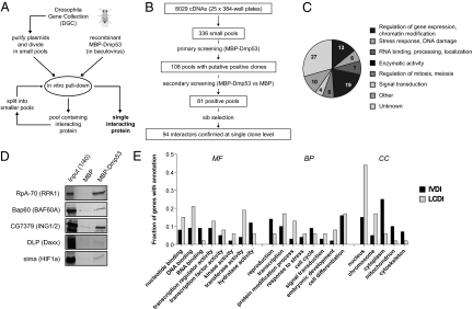
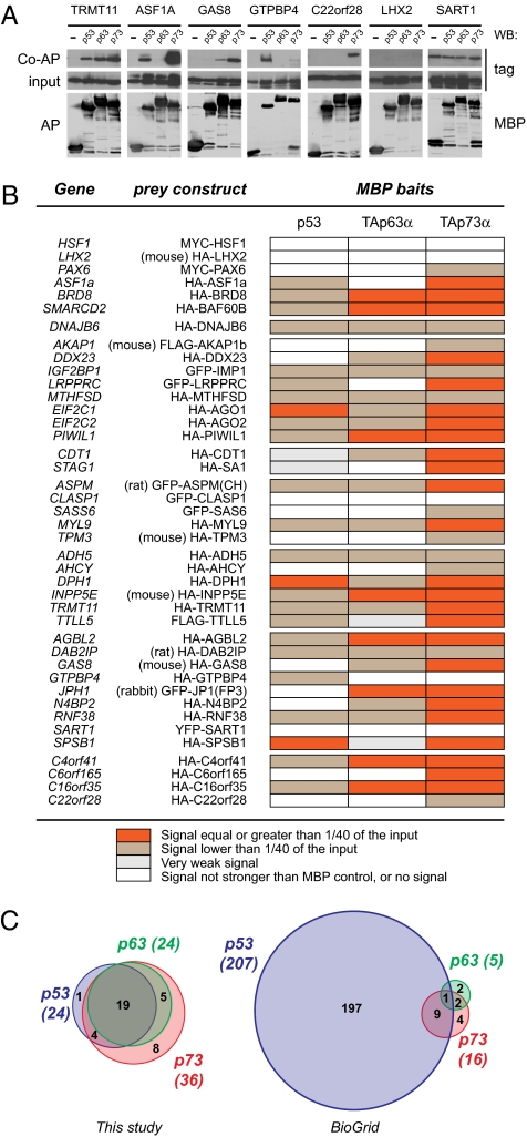
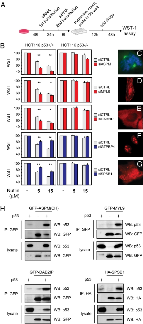
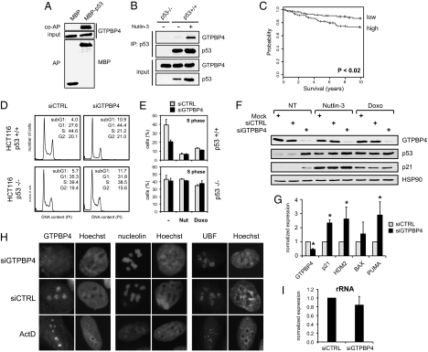
The genome of the fruitfly Drosophila melanogaster contains a single p53-like protein, phylogenetically related to the ancestor of the mammalian p53 family of tumor suppressors. We reasoned that a comprehensive map of the protein interaction profile of Drosophila p53 (Dmp53) might help identify conserved interactions of the entire p53 family in man. Using a genome-scale in vitro expression cloning approach, we identified 91 previously unreported Dmp53 interactors, considerably expanding the current Drosophila p53 interactome. Looking for evolutionary conservation of these interactions, we tested 41 mammalian orthologs and found that 37 bound to one or more p53-family members when overexpressed in human cells. An RNAi-based functional assay for modulation of the p53 pathway returned five positive hits, validating the biological relevance of these interactions. One p53 interactor is GTPBP4, a nucleolar protein involved in 60S ribosome biogenesis. We demonstrate that GTPBP4 knockdown induces p53 accumulation and activation in the absence of nucleolar disruption. In breast tumors with wild-type p53, increased expression of GTPBP4 correlates with reduced patient survival, emphasizing a potential relevance of this regulatory axis in cancer.


Conflict of interest statement

The authors declare no conflict of interest.

Figures

Fig. 1.
Fig. 1.
Small-pool IVEC screen for Drosophila p53 interactors. (A) Schematic diagram of the small-pool Drosophila IVEC approach used to identify Dmp53 binding proteins. (B) Flow chart summarizing data relative to each step of the screening. (C) Pie chart describing 92 identified in vitro Dmp53 interactors, arbitrarily divided into functional categories. (D) Identification of expected Dmp53 interactors. Shown are in vitro pull-down experiments relative to five proteins whose human orthologs (in parentheses) are known interactors of p53. (E) Comparison of Dmp53 interactors identified in this screening (IVDI, in vitro Dmp53 interactors) with Dmp53 interactors reported in previous studies (LCDI, literature-curated Dmp53 interactors). The fractions of proteins with the indicated GO slim annotations in the IVDI (solid bars) and LCDI (shaded bars) datasets are shown. The histogram reports only the most abundant (>5%) GO slim annotations in the categories of molecular function (MF), biological process (BP), and cell compartment (CC).
Fig. 2.
Fig. 2.
Binding of human orthologs of Dmp53 interactors to p53 and p53-related proteins. (A) Seven representative co-AP experiments. Shown are examples of preys that interacted with all three p53-family proteins (TRMT11), preys that bound to selected members of the p53 family (ASF1A, GAS8, GTPBP4, and C22orf28), and preys that were scored as not interacting (LHX2 and SART1). (Top) Tagged prey proteins after coaffinity purification. (Middle) Expression of prey in total lysates (1/40th of the input). (Bottom) MBP-tagged baits after affinity purification. The Top and Middle are cropped from the same autoradiography (i.e., have the same exposure). (B) Table summarizing the results of co-AP assays for 41 mammalian orthologs of Dmp53 interacting proteins. Expression plasmids encoding the indicated tagged proteins were cotransfected with pcDNA3-MBP, pcDNA3-MBP-p53, pcDNA3-MBP-TAp63α, or pcDNA3-MBP-TAp73α in 293T cells. MBP fusion proteins (baits) were purified on amylose beads, and copurified proteins were detected by immunoblotting. Strength of interaction was scored according to the fraction of the input prey protein that copurified with MBP baits. (C) Venn diagrams summarizing protein interactions associated with p53 family members in mammals. The results of this study are compared to current data available in protein–protein interaction databases (BioGrid release 2.0.45).
Fig. 3.
Fig. 3.
Functional validation of p53 interactors by RNAi. (A) Schematic diagram of the experimental procedure. p53-dependent growth inhibition was assayed after knockdown of newly identified potential p53 interactants. After siRNA transfection, cells were trypsinized and counted, and identical numbers of cells were seeded in 96-well plates. Drugs were added 12 h later, and WST-1 activity was measured after an additional 48 h. (B) Transient knockdown of five putative p53 interactors affects the response of HCT116 WT cells to Nutlin-3. To control p53 dependency, the same experiment was done in HCT116 p53−/− cells (*, P < 0.05; **, P < 0.01; n = 3). (CG) Cellular localization of functionally validated p53 interactors. C shows localization of endogenous ASPM in a mitotic U2OS cell, with Hoechst counterstaining for metaphase chromosomes. D–G show immunofluorescence of the indicated HA-tagged proteins transiently transfected in U2OS cells. (H) Coimmunoprecipitation of p53 with four functionally validated interactors. Expression plasmids encoding the indicated proteins were cotransfected in p53-null H1299 cells. After cell lysis, tagged proteins were immunoprecipitated using antibodies cross-linked to protein G beads and analyzed by immunoblotting. Expression of transfected proteins in the lysate (1/40th of the input) is shown in the Lower part.
Fig. 4.
Fig. 4.
Evidence of a functional link between the nucleolar protein GTPBP4 and p53. (A) Exogenous p53 binds endogenous GTPBP4. Coaffinity purification of GTPBP4 with overexpressed MBP-p53 in U2OS cells is shown. (Top) Endogenous GTPBP4 after coaffinity purification. (Middle) Expression of GTPBP4 in total lysates (1/40th of the input). (Bottom) MBP and MBP-p53 proteins after affinity purification. (B) Interaction between endogenous GTPBP4 and p53. Coimmunoprecipitation is shown of GTPBP4 with p53 in HCT116 WT cells untreated or treated for 12 h with 5 μM Nutlin-3. (Upper) Proteins immunoprecipitated using the anti-p53 monoclonal antibody (DO-1). (Lower) Endogenous p53 and GTPBP4 proteins in the lysates (1/40th of the input). As negative control, immunoprecipitation was performed in HCT116 p53−/− cells. (C) Cumulative survival (Kaplan–Meier) curves showing that higher expression of GTPBP4 correlates with reduced survival in breast cancer bearing wild-type p53 (30). Probesets corresponding to GTPBP4 were averaged and divided into low expressers (88 samples) and high expressers (93 samples), using the median GTPBP4 expression as cutoff. The two groups display a significant difference in survival rates (P = 0.0193). (D) Transient knockdown of GTPBP4 induces p53-dependent inhibition of cell proliferation. FACS analysis is shown of HCT116 WT and HCT116 p53−/− cells transfected with control or GTPBP4 siRNA. (E) Knockdown of GTPBP4 reduces S-phase in cells bearing p53. Average S-phase fraction is shown of HCT116 WT and HCT116 p53−/− cells transfected with control or GTPBP4 siRNA and treated for 24 h with 5 μM Nutlin-3 or 0.05 μM Doxorubicin. Values are means ± SD (n = 3). (F) Knockdown of GTPBP4 induces accumulation of p53 and p21 proteins. Immunoblotting is shown of HCT116 WT cells transfected with control or GTPBP4 siRNA and left untreated (NT) or treated for 24 h with Nutlin-3 (5 μM) or Doxorubicin (0.05 μM). A specific antibody to GTPBP4 confirmed efficient knockdown of the endogenous protein. Hsp90 was detected in the same lysates as a loading control. (G) Knockdown of GTPBP4 promotes transcription of p53-target genes. RT-qPCR shows up-regulation of p53-target genes in HCT116 WT cells transfected for 48 h with GTPBP4 siRNA. mRNA expression is normalized to GAPDH. Analysis of GTPBP4 mRNA in the same samples confirms the efficiency of knockdown (*, P < 0.05; n = 3). (H) GTPBP4 knockdown does not affect localization of key nucleolar proteins. Immunofluorescence analysis is shown of GTPBP4, Nucleolin, and UBF in U2OS cells transfected for 48 h with either control or GTPBP4 siRNA. The same proteins were analyzed in U2OS cells treated with 5 nM Actinomycin D for 12 h. Nuclei are counterstained with Hoechst. (I) GTPBP4 knockdown does not affect rRNA transcription. HCT116 WT cells were transfected with control or GTPBP4 siRNA, and prerRNA levels were quantified by RT-qPCR after 48 h, using primers specific for the 5′ external transcribed spacer of pre-rRNA and normalizing for GAPDH. Values are means ± SD.

Similar articles

Cited by

References

    1. Vousden KH, Lu X. Live or let die: The cell's response to p53. Nat Rev Cancer. 2002;2:594–604. - PubMed
    1. Hainaut P, et al. Database of p53 gene somatic mutations in human tumors and cell lines: Updated compilation and future prospects. Nucleic Acids Res. 1997;25:151–157. - PMC - PubMed
    1. Mantovani F, et al. The prolyl isomerase Pin1 orchestrates p53 acetylation and dissociation from the apoptosis inhibitor iASPP. Nat Struct Mol Biol. 2007;14:912–920. - PubMed
    1. Marchenko ND, Wolff S, Erster S, Becker K, Moll UM. Monoubiquitylation promotes mitochondrial p53 translocation. EMBO J. 2007;26:923–934. - PMC - PubMed
    1. Kruse J-P, Gu W. Modes of p53 regulation. Cell. 2009;137:609–622. - PMC - PubMed

Publication types