. 2010 Apr 6;107(14):6364-9.
doi: 10.1073/pnas.0915063107. Epub 2010 Mar 22.

Origin and formation of the first two distinct cell types of the inner cell mass in the mouse embryo

Affiliations

Origin and formation of the first two distinct cell types of the inner cell mass in the mouse embryo

Samantha A Morris et al. Proc Natl Acad Sci U S A. .

Abstract

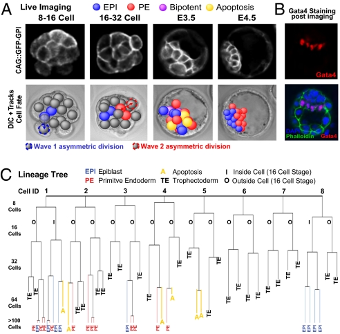
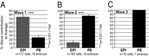
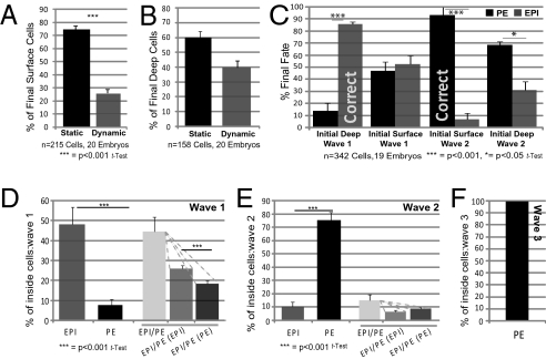
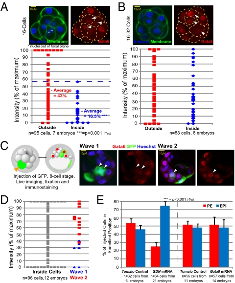
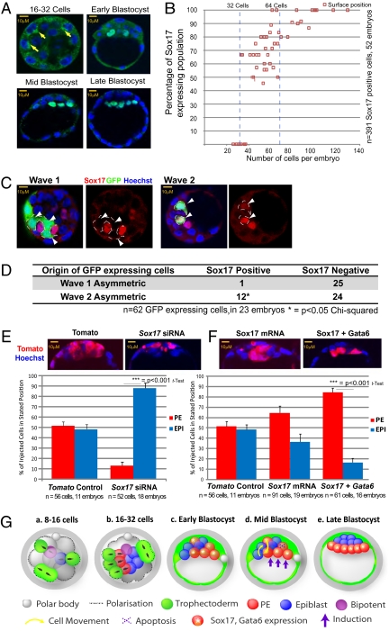
A crucial question in mammalian development is how cells of the early embryo differentiate into distinct cell types. The first decision is taken when cells undertake waves of asymmetric division that generate one daughter on the inside and one on the outside of the embryo. After this division, some cells on the inside remain pluripotent and give rise to the epiblast, and hence the future body, whereas others develop into the primitive endoderm, an extraembryonic tissue. How the fate of these inside cells is decided is unknown: Is the process random, or is it related to their developmental origins? To address this question, we traced all cells by live-cell imaging in intact, unmanipulated embryos until the epiblast and primitive endoderm became distinct. This analysis revealed that inner cell mass (ICM) cells have unrestricted developmental potential. However, cells internalized by the first wave of asymmetric divisions are biased toward forming pluripotent epiblast, whereas cells internalized in the next two waves of divisions are strongly biased toward forming primitive endoderm. Moreover, we show that cells internalized by the second wave up-regulate expression of Gata6 and Sox17, and changing the expression of these genes determines whether the cells become primitive endoderm. Finally, with our ability to determine the origin of cells, we find that inside cells that are mispositioned when they are born can sort into the correct layer. In conclusion, we propose a model in which the timing of cell internalization, cell position, and cell sorting combine to determine distinct lineages of the preimplantation mouse embryo.


Conflict of interest statement

The authors declare no conflict of interest.

Figures

Fig. 1.
Fig. 1.
Live cell imaging and tracking. (A) GFP-GPI expression from E.2.5 to E4.5. Deconvolved fluorescence and differential interference contrast time-lapse images overlaid with Simi BioCell cell-tracking spheres. (B) Embryos stained to reveal Gata4-positive cells adjacent to mature blastocyst cavity confirming normal development during each imaging session. Gata4-positive cells were present in a one-cell-thick surface layer. (C) Lineage tree from representative embryo. All cells were traced to the early 32-cell blastocyst; then inside cells were traced to late blastocyst. A cell was defined as occupying an inside position by the orientation of the cell division that generated it, by its enclosure from the outside environment by neighboring cells, and by continued analysis of its position as development progressed. Allocation to trophectoderm (TE), EPI, or PE and apoptosis (A) are indicated.
Fig. 2.
Fig. 2.
Proportion of the first (A), second (B), and third (C) waves of asymmetric division contributing to EPI and PE.
Fig. 3.
Fig. 3.
Cell movement and fate in the ICM. ICM surface (A) and deep (B) cell dynamics from early to late blastocyst. (C) Relationship of final cell fate to cell origin and behavior. Cells are grouped according to final fate and are classified according to originating asymmetric division and initial ICM position. Cells from wave 2 initially at the ICM surface are highly likely to become PE (>90%, P < 0.001, t test). Cells from wave 1 initially deep contribute significantly to EPI (almost 90%, P < 0.001, t test). “Correct” refers to allocation of wave 1 cells to EPI and wave 2 cells to PE before cell movement; n = 19, because one embryo was analyzed from the 32-cell stage. (DF) Bipotency in wave 1 (D), in wave 2 (E), and in wave 3 (F) showing mean values of proportions of cells per embryo with indicated final fates.
Fig. 4.
Fig. 4.
Gata6 expression and PE formation. Immunostaining of Gata6 in (A) 16-cell and (B) 16- to 32-cell embryos. Gata6 staining intensities are expressed as a percentage of maximum. White arrowheads indicate inside cells. (C) Correlation of Gata6 expression with wave of origin. Shown is an eight-cell blastomere injected with GFP mRNA, monitored for wave 1 or wave 2 division and immunostained for Gata6 at E3.5. White arrowheads indicate inside cells expressing GFP. (D) Gata6 levels in inside cells, relative to cells from a specific wave. Intensities are expressed as a percentage of maximum. (E) Cells injected with dominant negative Gata6 (GDN) localized deep in the ICM in 74.8% of cases (P < 0.001, t test, n = 64 cells from 21 embryos). Overexpression of Gata6 in eight-cell blastomeres does not alter cell fate.
Fig. 5.
Fig. 5.
Sox17 expression and PE formation. (A) Staining to reveal Sox17 (yellow arrows indicate cytoplasmic staining) from 16- to 32-cell embryos to E4.5. (B) Proportion of cells in surface ICM with nuclear Sox17 as development proceeds. (C) Correlation of Sox17 expression with wave of origin. An eight-cell blastomere was injected with GFP mRNA and associated with a particular division wave before staining for Sox17 at the 32-cell stage. White arrowheads indicate inside cells expressing GFP. (D) Origins of Sox17-positive and -negative cells in relation to division waves from C. Sox17 expression was seen in 92% of GFP-expressing cells from wave 2, compared with 12% of such cells from wave 1 (P > 0.05, χ2 test). (E) Sox17 RNAi directs cellular descendents to EPI rather than PE. (F) Cells overexpressing Sox17 and Gata6 are directed preferentially to PE. (G) Working model. (a and b) Cells generated in the first wave of asymmetric division are biased to generate EPI and bipotent precursors, whereas cells generated in the subsequent waves are biased to generate PE over EPI. Black arrows indicate orientation of cell division. (c) PE progenitors express Sox17 and higher levels of Gata6. Most inner cells are positioned according to their fate when the embryo cavitates in deep or surface ICM, but some are not. (d) Cells positioned within an inappropriate layer tend to relocate according to their wave of origin and fate: PE-destined cells relocate to the surface, and EPI-destined cells relocate deep. Some cells, particularly in deep ICM, apoptose. Hypothetically, induction from the cavity might further enhance PE fate in surface cells. (e) In the mature blastocyst, PE is fully segregated from EPI.

Similar articles

Cited by

References

    1. Johnson MH, Ziomek CA. The foundation of two distinct cell lineages within the mouse morula. Cell. 1981;24:71–80. - PubMed
    1. Barlow P, Owen DAJ, Graham CF. DNA synthesis in the preimplantation mouse embryo. J Embryol Exp Morphol. 1972;27:431–445. - PubMed
    1. Pedersen RA, Wu K, Bałakier H. Origin of the inner cell mass in mouse embryos: Cell lineage analysis by microinjection. Dev Biol. 1986;117:581–595. - PubMed
    1. Fleming TP. A quantitative analysis of cell allocation to trophectoderm and inner cell mass in the mouse blastocyst. Dev Biol. 1987;119:520–531. - PubMed
    1. Dyce J, George M, Goodall H, Fleming TP. Do trophectoderm and inner cell mass cells in the mouse blastocyst maintain discrete lineages? Development. 1987;100:685–698. - PubMed

Publication types