. 2010 Apr 6;107(14):6352-7.
doi: 10.1073/pnas.1000854107. Epub 2010 Mar 22.

Xenopus Bsx links daily cell cycle rhythms and pineal photoreceptor fate

Affiliations

Xenopus Bsx links daily cell cycle rhythms and pineal photoreceptor fate

Silvia D'Autilia et al. Proc Natl Acad Sci U S A. .

Abstract

In the developing central nervous system, the cell cycle clock plays a crucial role in determining cell fate specification. A second clock, the circadian oscillator, generates daily rhythms of cell cycle progression. Although these two clocks interact, the mechanisms linking circadian cell cycle progression and cell fate determination are still poorly understood. A convenient system to address this issue is the pineal organ of lower vertebrates, which contains only two neuronal types, photoreceptors and projection neurons. In particular, photoreceptors constitute the core of the pineal circadian system, being able to transduce daily light inputs into the rhythmical production of melatonin. However, the genetic program leading to photoreceptor fate largely remains to be deciphered. Here, we report a previously undescribed function for the homeobox gene Bsx in controlling pineal proliferation and photoreceptor fate in Xenopus. We show that Xenopus Bsx (Xbsx) is expressed rhythmically in postmitotic photoreceptor precursors, reaching a peak during the night, with a cycle that is complementary to the daily rhythms of S-phase entry displayed by pineal cells. Xbsx knockdown results in increased night levels of pineal proliferation, whereas activation of a GR-Xbsx protein flattens the daily rhythms of S-phase entry to the lowest level. Furthermore, evidence is presented that Xbsx is necessary and sufficient to promote a photoreceptor fate. Altogether, these data indicate that Xbsx plays a dual role in contributing to shape the profile of the circadian cell cycle progression and in the specification of pineal photoreceptors, thus acting as a unique link between these two events.


Conflict of interest statement

The authors declare no conflict of interest.

Figures
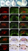
Fig. 1.
Fig. 1.
Xbsx expression during pineal organ development. Xbsx whole-mount in situ hybridization was performed at stage 16 (A), stage 19 (B), stage 22 (C), stage 32 (D), and stage 38 (E). Immunostaining for BrdU incorporation (G, J, M, and P; green staining), in situ hybridization for Xbsx (F, I, L, and O; red staining), and merge (H, K, N, and Q) were performed on cryostat sections at stage 24 (FH), stage 32 (IK), stage 37 (LN), and stage 40 (OQ). In situ hybridization for Xbsx (R, red staining), Xotx5 (S, blue staining), and merge (T) was performed on stage 37 cryostat sections. In situ hybridization for Xbsx (U, red staining), Hermes (V, blue staining), and merge (W) was performed on stage 37 cryostat sections.
Fig. 2.
Fig. 2.
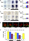
Daily rhythms of Xbsx expression and S-phase entry of pineal cells. (A and C) Whole-mount in situ hybridization for Xbsx and Tph was performed on dissected stage 46 brains. Embryos were kept in an LD (A) or DD (C) cycle and collected at the indicated time points (ZT0, lights on; ZT12, lights off). Quantification was determined by optical density (O.D.) of Xbsx and Tph in situ hybridization signal for embryos kept in LD (B) or DD (D) condition. The peak-to-trough statistical difference was determined by the Student's t test: Xbsx, P = 0.006; Tph, P = 0.002 (B) and Xbsx, P = 0.21; Tph, P = 0.008 (D). (E) Representative cryostat sections showing BrdU incorporation (green staining) in the pineal organ at five time points between stage 26 and stage 36. The Xotx5-positive area (red staining) is circled. (F) Quantification of BrdU-labeled cells present in the Xotx5-positive area. At each time point, the mean percentage per pineal organ of BrdU-positive nuclei over the total number of nuclei was plotted against ZT time. The embryonic stage (St.) corresponding to each time point is indicated on the top. Data represent pooled results from three independent experiments. The peak-to-trough statistical difference was determined by the Student's t test: P = 0.001. The number of cells counted is shown in Table S1. In this and the following figures, the white and black bars indicate the light and dark phases, respectively. The gray bar in DD experiments indicates a dark phase at the time when control embryos are in the light phase.
Fig. 3.
Fig. 3.
Xbsx knockdown leads to increased S-phase entry during the night and to a reduction of pineal photoreceptors. (A) Representative cryostat sections showing BrdU incorporation (green staining) in control- and MoXbsx-injected embryos during 24 h between stage 26 and stage 34. The Xotx5-positive area (red staining) is circled. (B) Quantification of BrdU-labeled cells present in the Xotx5-positive area of control-injected (blue bars) and MoXbsx-injected (red bars) embryos at eight time points. At each time point, the mean percentage per pineal organ of BrdU-positive nuclei over the total number of nuclei is plotted against ZT time. Data represent pooled results from three independent experiments. The number of cells counted is shown in Table S1. (C) Average number of TUNEL-positive nuclei in the Xotx5-positive area per pineal organ for embryos collected at ZT6 and ZT18, corresponding to stage 36 and stage 38, respectively. Data represent pooled results from three independent experiments. Six control (Co)- and six MoXbsx-injected embryos were analyzed per time point in each experiment. (D and E) Analysis of pineal cell types in control- and MoXbsx-injected stage 42 tadpoles. (D) Representative cryostat sections showing expression of markers for photoreceptors (Recoverin) and projection neurons (Hermes). (E) Average numbers of cells positive for Recoverin and Hermes per pineal organ. Data represent pooled results from three independent experiments. For each marker, four control (Co)-injected and four MoXbsx-injected embryos were analyzed per time point in each experiment. Asterisks indicate statistical differences as determined by the Student's t test: **P < 0.01; ***P < 0.001. Error bars indicate SEM.
Fig. 4.
Fig. 4.
Overexpression of Xbsx reduces S-phase entry and increases the number of pineal photoreceptors. (A) Representative cryostat sections showing BrdU incorporation (green staining) at four time points in embryos injected with GR-Xbsx and treated or untreated with dexamethasone (DEX). The Xotx5-positive area (red staining) is circled. (B) Quantification of BrdU-labeled cells present in the Xotx5-positive area of embryos injected with GR-Xbsx and untreated (gray bars) or treated (green bars) with DEX. At each time point, the mean percentage per pineal organ of BrdU-positive nuclei over the total number of nuclei is plotted against ZT time. Data represent pooled results from three independent experiments. The number of cells counted is shown in Table S1. **P < 0.01; ***P < 0.001. (C) Average number of TUNEL-positive nuclei in the Xotx5-positive area per pineal organ for embryos collected at ZT6 and ZT18, corresponding to stage 36 and stage 38, respectively. Data represent pooled results from three independent experiments. Six DEX-treated and six untreated GR-Xbsx-injected embryos were analyzed per time point in each experiment. (D and E) Analysis of pineal cell types in stage 42 tadpoles. Embryos were injected with GR-Xbsx and allowed to develop in the presence or absence of DEX. (D) Representative cryostat sections showing expression of markers for photoreceptors (Recoverin) and projection neurons (Hermes). (E) Average numbers of cells positive for Recoverin and Hermes per pineal organ. Data represent pooled results from three independent experiments. For each marker, four DEX-treated and four untreated GR-Xbsx-injected embryos were analyzed per time point in each experiment. Asterisks indicate statistical differences as determined by the Student's t test: **P < 0.01. Error bars indicate SEM. (F) RT-PCR analysis to evaluate the effects of cyclin A2/cdk2 overexpression on Xbsx expression. Control and injected embryos were collected at the indicated time points. ODC, ornithine decarboxylase.
Fig. 5.
Fig. 5.
Working model for Xbsx function in cell proliferation and differentiation of photoreceptor precursors. (A) Schematic summary of the relation between Xbsx expression and pineal cell proliferation in WT and manipulated embryos. (B) Model of action for Xbsx. The pink cell represents the common progenitor of projection neurons and photoreceptors. It is assumed that photoreceptor precursors undergo a limited number of asymmetrical cell divisions. Xbsx knockdown prevents cell cycle exit of photoreceptor precursors that eventually undergo apoptosis. Xbsx overexpression increases cell cycle exit of photoreceptor precursors and promotes their differentiation.

Similar articles

Cited by

References

    1. Ohnuma S, Harris WA. Neurogenesis and the cell cycle. Neuron. 2003;40:199–208. - PubMed
    1. Andreazzoli M. Molecular regulation of vertebrate retina cell fate. Birth Defects Res., Part C. 2009;87:284–295. - PubMed
    1. Okamura H. Clock genes in cell clocks: Roles, actions, and mysteries. J Biol Rhythms. 2004;19:388–399. - PubMed
    1. Hunt T, Sassone-Corsi P. Riding tandem: Circadian clocks and the cell cycle. Cell. 2007;129:461–464. - PubMed
    1. Roenneberg T, Foster RG. Twilight times: Light and the circadian system. Photochem Photobiol. 1997;66:549–561. - PubMed

Publication types