. 2010 Apr 6;107(14):6346-51.
doi: 10.1073/pnas.1002401107. Epub 2010 Mar 22.

Coordination of Rab8 and Rab11 in primary ciliogenesis

Affiliations

Coordination of Rab8 and Rab11 in primary ciliogenesis

Andreas Knödler et al. Proc Natl Acad Sci U S A. .

Abstract

Primary cilia are microtubule-based membrane projections located at the surface of many cells. Defects in primary cilia formation have been implicated in a number of genetic disorders, such as Bardet-Biedl Syndrome and Polycystic Kidney Disease. Recent studies have demonstrated that polarized vesicular transport involving Rab8 and its guanine nucleotide-exchange factor Rabin8 is essential for primary ciliogenesis. Here we report that Rabin8 is a direct downstream effector of Rab11, which functions in membrane trafficking from the trans-Golgi network and recycling endosomes. Rab11, in its GTP-bound form, interacts with Rabin8 and kinetically stimulates the guanine nucleotide-exchange activity of Rabin8 toward Rab8. Rab11 is enriched at the base of the primary cilia and inhibition of Rab11 function by a dominant-negative mutant or RNA interference blocks primary ciliogenesis. Our results suggest that Rab GTPases coordinate with each other in the regulation of vesicular trafficking during primary ciliogenesis.


Conflict of interest statement

The authors declare no conflict of interest.

Figures

Fig. 1.
Fig. 1.
Rabin8 interacts with Rab11. (A) Binding of Hisx6-tagged wild-type and mutant Rab11a to GST-Rabin8. Rab11a preferentially bound to Rabin8 in the presence of GTPγS versus GTPβS. As a control, Rabin8 did not bind to Rab5a. Rab11a was detected by the anti-Hisx6 antibody. (B) Rabin8 bound to Rab11a[Q70L] stronger than to wild-type Rab11a. Rab11a[S25N] barely bound to Rabin8. (C) Diagram of GST fusion constructs of Rabin8 with serial C-terminal deletions. Numbers indicate the amino acid sequences. The GEF domain (amino acids 144–245) on Rabin8 is marked in black. (D) Coomassie Blue-stained gel showing the different Rabin8 fusion proteins coupled to Glutathione Sepharose. GST alone was used as a control. Rab11a was expressed as a Hisx6-tagged fusion protein. Binding of Rab11 to the individual Rabin8 constructs was detected with a monoclonal antibody against the Hisx6 epitope.
Fig. 2.
Fig. 2.
Rab11 stimulates the GEF activity of Rabin8. (A) Coomassie Blue-stained SDS/PAGE showing purified Rabin8, Rab8, and Trx-Hisx6-S-tagged Rab11a[Q70L], Rab5a[Q79L], Rab3a[Q81L]. Molecular weight (MW) is indicated to the left. The asterisks indicate the positions of Rabin8 cleaved from the GST tag and Rab8 cleaved from the NusA-Hisx6 tag, respectively. The additional bands present in the purification are most likely the degradation products of Rabin8 or Rab8, based on their recognition by the anti-Rabin8 or Rab8 antibodies (see Fig. S1 for details). Trx-Hisx6-S-tagged Rab11a[Q70L], Rab5a[Q79L], and Rab3a[Q81L] have the predicted molecular weights, as they are fusions of the Rab proteins with the 18 kDa Trx-Hisx6-S tag. (B) The release of [3H]GDP from Rab8 catalyzed by Rabin8 in the presence and absence of Rab11a[Q70L] was analyzed as described in Materials and Methods. Rab11a[Q70L] stimulated the release of [3H]GDP from Rab8 in the presence of Rabin8 (red). Rab11a[Q70L] significantly enhanced the GEF activity of Rabin8 toward Rab8 (P < 0.01, n = 3). (C) Rabin8-mediated [3H]GDP release from Rab8 was tested in the absence (black bars) and presence (white bars) of Rab5a[Q79L] or Rab3a[Q81L] at 5 min and 20 min (n = 3). Rab5a[Q79L] or Rab3a[Q81L] did not stimulate Rabin8-mediated release of [3H]GDP from Rab8. The data were analyzed using Student's t test and presented as SEM (n = 3).
Fig. 3.
Fig. 3.
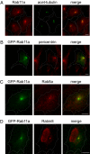
Localization of Rab11 at the base of the primary cilia. The localization of endogenous Rab11a (A) and expressed GFP-Rab11a (B–D) was examined in hTERT-RPE1 cells together with the cilia marker acetylated tubulin (A, green); the basal body marker pericentrin (B, red), the cilia membrane marker Rab8a (C, red). Rabin8 was stained with the affinity-purified anti-Rabin8 antibody as previously described (10) (D, red). Merged images are shown to the right. (Scale bars, 10 μm.) Cell boundaries are outlined.
Fig. 4.
Fig. 4.
Inhibition of Rab11 function in cells blocks primary ciliogenesis. (A) hTERT-RPE1 cells were transfected with the dominant-negative Rab11a[S25N] and GFP-Rab5a[S34N] mutants. The length of primary cilia in these cells was determined by immunostaining of acetylated α-tubulin (red). GFP-Rab11a[S25N]-expressing cells (green) had much shorter cilia than the untransfected cells in the same field. As a control, cells transfected with GFP-Rab5a[S34N] had normal cilia. Nuclei were stained with DAPI (blue) and the merged images are shown to the right. (Scale bar, 10 μm.) (B) Quantification of cilia length in untransfected, GFP-Rab11a[S25N]- and GFP-Rab5a[S34N]-expressing cells. The data were analyzed using Student's t test and presented as SEM (n = 50, P < 0.01) when comparing GFP-Rab11a[S25N]-expressing cells with the other groups. (C) hTERT-RPE1 cells were treated with siRNA oligos targeting human Rab11a and Rab11b. Cells treated with siRNA oligos against Luciferase (LUC) were used as controls. For the rescue experiment, the Rab11 knockdown cells were transfected with a GFP-Rab11a variant (RAB11’) with nucleotide mutations that mismatch the Rab11 oligos (SI Materials and Methods). Rab11 levels were analyzed by Western blot using the anti-Rab11 polyclonal antibody. (D) The primary cilia in Rab11 knockdown cells were significantly shorter than those in LUC siRNA control cells. Cells transfected with GFP-Rab11a' variant (RAB1' Rescue, green) have near normal length cilia. Primary cilia were immunostained with a mouse monoclonal antibody against acetylated α-tubulin (red). The nuclei were stained with DAPI (blue). The merged images are shown to the right. (Scale bar, 10 μm.) (E) Quantification of cilia length in different cells. The data were analyzed using Student's t test and presented as SEM, n = 60, P < 0.01.
Fig. 5.
Fig. 5.
Effect of Rab11 on Rabin8-BBS1 interaction. (A) Mapping the domain of Rabin8 that binds to BBS1. Cell lysates from hTERT-RPE1 cells expressing myc-BBS1 were incubated with GST or GST-Rabin8 fusion proteins with serial truncations at the C terminus (Upper, Ponceau S. staining). The bound myc-BBS1 was analyzed by Western blot using the anti-myc monoclonal antibody. Myc-BBS1 interacts with full-length Rabin8, Rabin8 (amino acid 1–400) and Rabin8 (amino acid 1–331). Further deletion of the C terminus abolished this interaction. Molecular weights (MW, in kilodaltons) are indicated to the right. (B) Expression of activated Rab11 promotes the association of BBS1 with Rabin8. hTERT-RPE1 cells were cotransfected with pEGFP-C1, pEGFPC1-myc-Rab11a, pEGFPC1-myc-Rab11a[Q70L], or pEGFPC1-myc-Rab11a[S25N] together with myc-BBS1. Two days after transfection, cells were harvested and immunoprecipitation assay was performed with anti-Rabin8 antibody. The amount of myc-BBS1 coimmunoprecipitated with Rabin8 was detected by Western blot using the anti-Myc monoclonal antibody. (Upper) The levels of myc-BBS1 and the GFP-myc-tagged Rab11 variants in the cell. (Lower) Myc-BBS1 coimmunoprecipitated by the Rabin8 antibody from the cell lysates. Myc-BBS1 binds to Rabin8 more strongly in cells expressing the Rab11a[Q70L] mutant.

References

    1. Satir P, Christensen ST. Overview of structure and function of mammalian cilia. Annu Rev Physiol. 2007;69:377–400. - PubMed
    1. Marshall WF. The cell biological basis of ciliary disease. J Cell Biol. 2008;180:17–21. - PMC - PubMed
    1. Gerdes JM, Davis EE, Katsanis N. The vertebrate primary cilium in development, homeostasis, and disease. Cell. 2009;137:32–45. - PMC - PubMed
    1. Leroux MR. Taking vesicular transport to the cilium. Cell. 2007;129:1041–1043. - PubMed
    1. Pazour GJ, Bloodgood RA. Targeting proteins to the ciliary membrane. Curr Top Dev Biol. 2008;85:115–149. - PubMed

Publication types

MeSH terms