. 2010 Apr 6;107(14):6358-63.
doi: 10.1073/pnas.0909998107. Epub 2010 Mar 22.

Estrogen receptor ESR1 controls cell migration by repressing chemokine receptor CXCR4 in the zebrafish posterior lateral line system

Affiliations

Estrogen receptor ESR1 controls cell migration by repressing chemokine receptor CXCR4 in the zebrafish posterior lateral line system

Laurent Gamba et al. Proc Natl Acad Sci U S A. .

Abstract

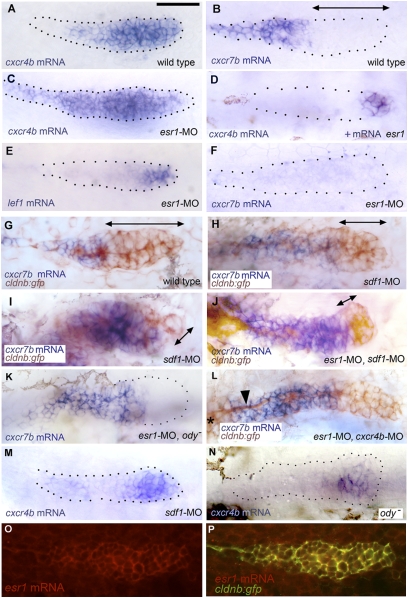
The primordium that generates the embryonic posterior lateral line of zebrafish migrates from the head to the tip of the tail along a trail of SDF1-producing cells. This migration critically depends on the presence of the SDF1 receptor CXCR4 in the leading region of the primordium and on the presence of a second SDF1 receptor, CXCR7, in the trailing region of the primordium. Here we show that inactivation of the estrogen receptor ESR1 results in ectopic expression of cxcr4b throughout the primordium, whereas ESR1 overexpression results in a reciprocal reduction in the domain of cxcr4b expression, suggesting that ESR1 acts as a repressor of cxcr4b. This finding could explain why estrogens significantly decrease the metastatic ability of ESR-positive breast cancer cells. ESR1 inactivation also leads to extinction of cxcr7b expression in the trailing cells of the migrating primordium; this effect is indirect, however, and due to the down-regulation of cxcr7b by ectopic SDF1/CXCR4 signaling in the trailing region. Both ESR1 inactivation and overexpression result in aborted migration, confirming the importance of this receptor in the control of SDF1-dependent migration.


Conflict of interest statement

The authors declare no conflict of interest.

Figures

Fig. 1.
Fig. 1.
The minimal promoter of cxcr4b. (A and B) transient expression of RFP under the control of the 139-bp cxcr4b promoter fragment in a migrating primordium (A) and in a deposited neuromast (B) in cldnb:gfp embryos. (Left) RFP fluorescence, (Right) RFP in red, merged with GFP in green. (C) Stable expression of RFP driven by the 139-bp promoter in a line doubly transgenic for cxcr4b:rfp and cldnb:gfp. (D) Coexpression of RFP (red) and GFP (green) in the same embryo. (C and D) Assembled from four consecutive frames of a Z-stack; the slight mismatch between green and red, in some cells, is due to migration during the imaging process. In this and all other figures anterior is to the left, and the primordium is migrating to the right. (Scale bars: 50 μm.) (E) Low-scale view of an embryo doubly transgenic for cxcr4b:rfp and cldnb:gfp, illustrating the complete absence of RFP expression in anterior lateral line neuromasts (ALL) and in the infraorbital anterior lateral line primordium (ALL io) as well as in the otic system (arrowheads). Faint RFP fluorescence can be detected in the PLL ganglion (PLLg). (F) Comparison of the sequences upstream of the TATA box in Danio cxcr4b, Homo cxcr4, and Danio cxcr4a promoters. The 139-bp cxcr4b minimal promoter defined by us begins one base upstream of the ESR1 putative binding site and includes the initiation ATG; the 157-bp minimal promoter defined in Homo (15) begins 10 bases upstream of the ESR1 binding site and does not include the initiation ATG. The corresponding sequence in the promoter of cxcr4a is also shown. Putative binding sites according the TESS software are colored. The ESR1 binding sites in cxcr4b and human cxcr4 (red) are both followed by a C, consistent with the presence of a pyrimidine at this position in the consensus binding site. The binding sites for SP1 and TATA Binding Protein have been shown to be instrumental for cxcr4 expression in the minimal human promoter (26).
Fig. 2.
Fig. 2.
Migration of the PLL primordium is altered in esr1-MO embryos. (A) Three frames of a time-lapse movie of primordium migration in a cldnb:gfp embryo at 35 (t = 0), 38, and 41 hpf, respectively. At 41 hpf, the primordium has just migrated out of the field. (B) Five frames of a time-lapse movie of the primordium in an esr1-MO1 injected embryo at 35 (t = 0), 38, 41, 44, and 48 hpf, respectively. Migration is two to three times slower than in untreated cldnb:gfp embryos and eventually comes to a halt around 45 hpf. (C) In the same embryo, the immobile primordium has fragmented into two neuromasts at 51 hpf. (Scale bar in A: 50 μm.)
Fig. 3.
Fig. 3.
Patterns of gene expression in the PLL primordium. (A and B) Expression of cxcr4b and cxcr7b in 32-hpf control embryos. (C and D) Expression of cxcr4b in conditions of esr1 loss-of-function (C) and gain-of-function (D). The change in expression between A and C was observed in 75% of the injected embryos, respectively, the change between A and D in 30% of the injected embryos. (E and F) Expression of lef1 and cxcr7b in esr1 loss-of-function conditions. (G and H) Expression of cxcr7b (blue) and of gfp (brown) in a 32-hpf control cldnb:gfp embryo (G), in an sdf1-MO embryo (H; an extreme case of cxcr7b domain expansion is shown in I). (J–L) Rescue of cxcr7b expression in esr1-MO embryos that are also sdf1-MO (J), mutant for ody (K), or morphant for cxcr4b (L; note the nerve, arrowhead, extending from the ganglion, asterisk at the left of the figure, under the stalled primordium). (M and N) Expression of cxcr4b in 32-hpf sdf1-MO injected (M) or ody (N) homozygous embryos; the photograph in N was taken under Nomarski optics to illustrate the extent of the primordium. (O and P) Expression of esr1 in a 32-hpf migrating primordium as revealed by fluorescent in situ hybridization using a rhodamine-coupled antidioxygenin antibody (O) and coexpression of esr1 and cldnb:gfp in the same embryo (P; confocal imaging). (A–F, K, and M) are bright-field pictures without Nomarski optics, to facilitate the detection of expression patterns. The primordia are outlined by dotted lines, based on images taken under Nomarski optics as illustrated in N. Double-headed arrows indicate the extent of the region where cxcr7b expression is not detectable.
Fig. 4.
Fig. 4.
Control of expression of cxcr4b and cxcr7b in the leading (Right) and trailing (Left) regions of the migrating primordium. The expression of cxcr4b, which is homogeneous before the onset of migration (7), is repressed in the trailing third by ESR1, and maintained in the leading two-thirds by positive feedback through SDF1/CXCR4 signaling. An additional activating mechanism, possibly under direct control of Wnt signaling, ensures expression of cxcr4b in the leading cells even in the absence of CXCR4 signaling. The complementary pattern of expression for cxcr7b reflects repression by SDF1/CXCR4 signaling and an additional repression by Wnt signaling in the leading cells (11). Single lines: controls at the level of gene expression; double lines: controls at the level of gene product activity. Black and gray indicate components that are active and inactive, respectively, in the leading and trailing regions. Dotted line indicates diffusion of FGF ligands induced in the leading cells by Wnt signaling (11). Altering FGF signaling also affects cxcr4b and cxcr7b expression (12), although it is not clear whether this effect is direct or is due to changes in Wnt signaling, and this level of control has therefore not been included in this tentative diagram. Question marks indicate plausible, but undemonstrated, interactions.

Similar articles

Cited by

References

    1. Ghysen A, Dambly-Chaudière C. The lateral line microcosmos. Genes Dev. 2007;21:2118–2130. - PubMed
    1. Metcalfe WK. Sensory neuron growth cones comigrate with posterior lateral line primordial cells in zebrafish. J Comp Neurol. 1985;238:218–224. - PubMed
    1. Gompel N, et al. Pattern formation in the lateral line of zebrafish. Mech Dev. 2001;105:69–77. - PubMed
    1. David NB, et al. Molecular basis of cell migration in the fish lateral line: Role of the chemokine receptor CXCR4 and of its ligand, SDF1. Proc Natl Acad Sci USA. 2002;99:16297–16302. - PMC - PubMed
    1. Li Q, Shirabe K, Kuwada JY. Chemokine signaling regulates sensory cell migration in zebrafish. Dev Biol. 2004;269:123–136. - PubMed

Publication types

MeSH terms

LinkOut - more resources