

Hypoxia inducible microRNA 210 attenuates keratinocyte proliferation and impairs closure in a murine model of ischemic wounds
- PMID: 20308562
- PMCID: PMC2872456
- DOI: 10.1073/pnas.1001653107
Hypoxia inducible microRNA 210 attenuates keratinocyte proliferation and impairs closure in a murine model of ischemic wounds
Abstract
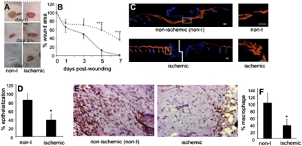
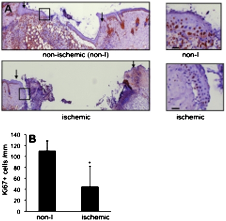
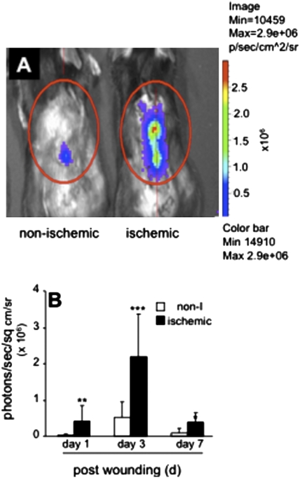
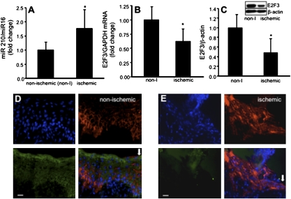
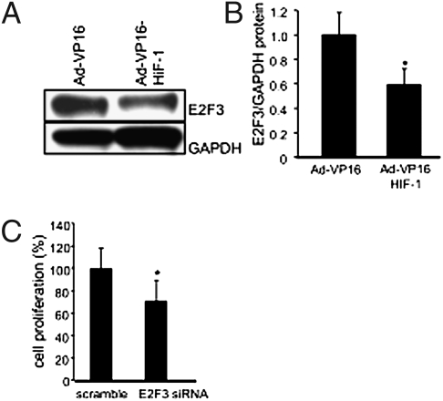
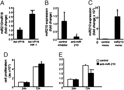
Ischemia complicates wound closure. Here, we are unique in presenting a murine ischemic wound model that is based on bipedicle flap approach. Using this model of ischemic wounds we have sought to elucidate how microRNAs may be implicated in limiting wound re-epithelialization under hypoxia, a major component of ischemia. Ischemia, evaluated by laser Doppler as well as hyperspectral imaging, limited blood flow and lowered tissue oxygen saturation. EPR oximetry demonstrated that the ischemic wound tissue had pO(2) <10 mm Hg. Ischemic wounds suffered from compromised macrophage recruitment and delayed wound epithelialization. Specifically, epithelial proliferation, as determined by Ki67 staining, was compromised. In vivo imaging showed massive hypoxia inducible factor-1alpha (HIF-1alpha) stabilization in ischemic wounds, where HIF-1alpha induced miR-210 expression that, in turn, silenced its target E2F3, which was markedly down-regulated in the wound-edge tissue of ischemic wounds. E2F3 was recognized as a key facilitator of cell proliferation. In keratinocytes, knock-down of E2F3 limited cell proliferation. Forced stabilization of HIF-1alpha using Ad-VP16- HIF-1alpha under normoxic conditions up-regulated miR-210 expression, down-regulated E2F3, and limited cell proliferation. Studies using cellular delivery of miR-210 antagomir and mimic demonstrated a key role of miR-210 in limiting keratinocyte proliferation. In summary, these results are unique in presenting evidence demonstrating that the hypoxia component of ischemia may limit wound re-epithelialization by stabilizing HIF-1alpha, which induces miR-210 expression, resulting in the down-regulation of the cell-cycle regulatory protein E2F3.
Conflict of interest statement
The authors declare no conflict of interest.
Figures
Similar articles
-
Acceleration of Diabetic Wound Healing with PHD2- and miR-210-Targeting Oligonucleotides.Tissue Eng Part A. 2019 Jan;25(1-2):44-54. doi: 10.1089/ten.TEA.2017.0484. Epub 2018 Jun 29. Tissue Eng Part A. 2019. PMID: 29644938 Free PMC article.
-
Fibroblast-Specific Deletion of Hypoxia Inducible Factor-1 Critically Impairs Murine Cutaneous Neovascularization and Wound Healing.Plast Reconstr Surg. 2015 Nov;136(5):1004-1013. doi: 10.1097/PRS.0000000000001699. Plast Reconstr Surg. 2015. PMID: 26505703 Free PMC article.
-
Polydeoxyribonucleotide restores blood flow in an experimental model of ischemic skin flaps.J Vasc Surg. 2012 Feb;55(2):479-88. doi: 10.1016/j.jvs.2011.07.083. Epub 2011 Nov 3. J Vasc Surg. 2012. PMID: 22051873
-
MiR equal than others: MicroRNA enhancement for cutaneous wound healing.J Cell Physiol. 2021 Dec;236(12):8050-8059. doi: 10.1002/jcp.30485. Epub 2021 Jun 23. J Cell Physiol. 2021. PMID: 34160067 Review.
-
MicroRNA-210: a unique and pleiotropic hypoxamir.Cell Cycle. 2010 Mar 15;9(6):1072-83. doi: 10.4161/cc.9.6.11006. Epub 2010 Mar 15. Cell Cycle. 2010. PMID: 20237418 Free PMC article. Review.
Cited by
-
Activation and regulation of the granulation tissue derived cells with stemness-related properties.Stem Cell Res Ther. 2015 Apr 29;6(1):85. doi: 10.1186/s13287-015-0070-9. Stem Cell Res Ther. 2015. PMID: 25925316 Free PMC article.
-
miR-136 modulates TGF-β1-induced proliferation arrest by targeting PPP2R2A in keratinocytes.Biomed Res Int. 2015;2015:453518. doi: 10.1155/2015/453518. Epub 2015 Jan 14. Biomed Res Int. 2015. PMID: 25654102 Free PMC article.
-
miR-210: the master hypoxamir.Microcirculation. 2012 Apr;19(3):215-23. doi: 10.1111/j.1549-8719.2011.00154.x. Microcirculation. 2012. PMID: 22171547 Free PMC article. Review.
-
microRNAs: innovative targets for cerebral ischemia and stroke.Curr Drug Targets. 2013 Jan 1;14(1):90-101. doi: 10.2174/138945013804806424. Curr Drug Targets. 2013. PMID: 23170800 Free PMC article. Review.
-
Elevated level of microRNA-210 at the initiation of muscular regeneration in acetic acid-induced non-ischemic skeletal muscular injury in mice.J Toxicol Pathol. 2022 Apr;35(2):183-192. doi: 10.1293/tox.2021-0061. Epub 2021 Dec 24. J Toxicol Pathol. 2022. PMID: 35516838 Free PMC article.
References
-
- Powell RJ, et al. Results of a double-blind, placebo-controlled study to assess the safety of intramuscular injection of hepatocyte growth factor plasmid to improve limb perfusion in patients with critical limb ischemia. Circulation. 2008;118:58–65. - PubMed
-
- Kerrigan CL, Daniel RK. Skin flap research: a candid view. Ann Plast Surg. 1984;13:383–387. - PubMed
Publication types
MeSH terms
Substances
Grants and funding
LinkOut - more resources
Full Text Sources
Other Literature Sources
Molecular Biology Databases
