. 2010 Apr 6;107(14):6510-5.
doi: 10.1073/pnas.0912315107. Epub 2010 Mar 22.

Zbtb20 is essential for the specification of CA1 field identity in the developing hippocampus

Affiliations

Zbtb20 is essential for the specification of CA1 field identity in the developing hippocampus

Zhifang Xie et al. Proc Natl Acad Sci U S A. .

Abstract

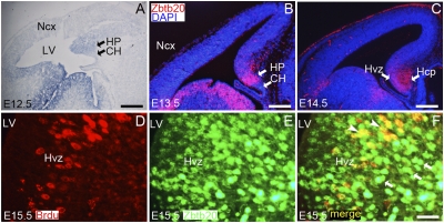
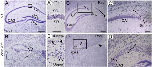
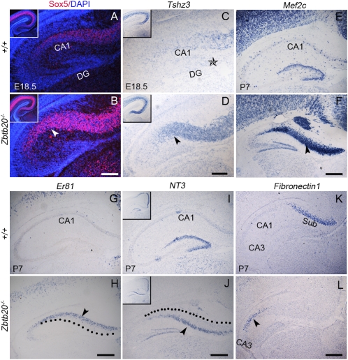
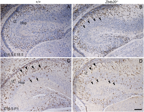
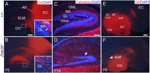
The development of hippocampal circuitry depends on the proper assembly of correctly specified and fully differentiated hippocampal neurons. Little is known about factors that control the hippocampal specification. Here, we show that zinc finger protein Zbtb20 is essential for the specification of hippocampal CA1 field identity. We found that Zbtb20 expression was initially activated in the hippocampal anlage at the onset of corticogenesis, and persisted in immature hippocampal neurons. Targeted deletion of Zbtb20 in mice did not compromise the progenitor proliferation in the hippocampal and adjacent transitional ventricular zone, but led to the transformation of the hippocampal CA1 field into a transitional neocortex-like structure, as evidenced by cytoarchitectural, neuronal migration, and gene expression phenotypes. Correspondingly, the subiculum was ectopically located adjacent to the CA3 in mutant. Although the field identities of the mutant CA3 and dentate gyrus (DG) were largely maintained, their projections were severely impaired. The hippocampus of Zbtb20 null mice was reduced in size, and exhibited increased apoptotic cell death during postnatal development. Our data establish an essential role of Zbtb20 in the specification of CA1 field identity by repressing adjacent transitional neocortex-specific fate determination.


Conflict of interest statement

The authors declare no conflict of interest.

Figures

Fig. 1.
Fig. 1.
Zbtb20 expression in the developing hippocampus. (A) Zbtb20 expression was examined by in situ hybridization on the sagittal section of E12.5 forebrain. (B and C) Zbtb20 expression was detected by immunohistochemistry using anti-Zbtb20 antibody 9A10 on the coronal sections through the forebrain at E13.5 (B) or E14.5 (C). (D–F) Double-immunostaining of anti-BrdU and anti-Zbtb20 on the E15.5 coronal brain sections. (F) The majority of BrdU+ cells in the hippocampal VZ (Hvz) were Zbtb20+ (yellow; indicated by arrowheads), and a large number of Zbtb20+ cells outside the Hvz were BrdU (green; indicated by arrows). HP, hippocampal primordium; Ncx, neocortex; Hcp, hippocampal cortical plate; LV, lateral ventricle. (Scale bars: 250 μm for A and C; 175 μm for B; 30 μm for D–F.)
Fig. 2.
Fig. 2.
Abnormal cytoarchitecture of the Zbtb20−/− hippocampus. (A–D) Nissl-stained coronal forebrain sections at P21. (A′ and B′) High-magnification views of the boxed areas in A and B, respectively. The normally single layer of the stratum pyramidale (SP; A′) was transformed into two principal layers in the mutant CA1 field (B′): an upper layer (small arrowheads) and a deep layer (large arrowheads). (C and D) At a more caudal level, a subiculum-like structure appeared adjacent to the mutant CA3. Dotted lines indicate boundaries of subiculum. (C′ and D′) High-magnification views of the boxed areas in C and D, respectively. Sub, subiculum. (Scale bars: 400 μm for A–D; 50 μm for A′ and B′; 100 μm for C′ and D′.)
Fig. 3.
Fig. 3.
Loss of hippocampus-specific markers in the Zbtb20−/− CA1 region. Coronal forebrain sections were detected for the indicated genes by in situ hybridization at the indicated stages. (A–F) NP2, EphA4, and EphA6 expression was present in the mutant CA3 and DG, but undetectable in the mutant CA1 (arrowheads in B, D, and F). (G and H) CA1-specific Man1α expression was totally lost in the mutant CA1 (arrowhead in H). (I–L) Decreased KA1 and Prox1 expression in the mutant CA3 (arrowhead in J) and DG (arrowhead in L), respectively. (Scale bars: 200 μm for A–F and I–L; 350 μm for G and H.)
Fig. 4.
Fig. 4.
Ectopic expression of transitional neocortex-specific genes in the Zbtb20−/− CA1 region. Coronal forebrain sections were detected for indicated genes by immunohistochemistry (A and B) or in situ hybridization (C–L) at the indicated stages. The arrowheads in B and D indicate the mutant CA1 field. The star in C indicates the notch at the DG crest. The arrowheads in H and J indicate the deep and upper layer of the mutant CA1, respectively. The dotted lines in H and J outline the upper and deep boundary of mutant CA1, respectively. (Insets) Low-magnification views of the hippocampus (A–D and I–J). (K and L) At caudal level, the expression domain of Fibronectin1, a marker for subiculum (K), was shifted adjacent to the mutant CA3 (L). (Scale bars: 200 μm for A–D; 250 μm for E–L.)
Fig. 5.
Fig. 5.
Accelerated migratory behavior of the Zbtb20−/− CA1 neurons. Hippocampal progenitors were labeled by BrdU incorporation at E16.5, and subjected to BrdU immunostaining at E18.5 or P1. The majority of wild-type CA1 neurons born at E16.5 (BrdU+ cells, dark brown) were still in the IZ (arrowheads in C) by P1 (A and C), whereas some of BrdU+ cells in the mutant CA1 had crossed the IZ and reached the CP by E18.5 (arrows in B), with the majority of them penetrating into the CP by P1 (arrows in D). (Scale bars: 175 μm.)
Fig. 6.
Fig. 6.
Impaired connections in Zbtb20−/− hippocampus. (A and B) Horizontal brain sections taken after the placement of DiI in the collateral EC at P5. (Insets) High-magnification views of the boxed areas in A and B, respectively. (C and D) Immunohistochemistry study for Calretinin-positive fibers on P14 saggital sections. (E and F) Schaffer collaterals projecting from the CA3 to the SO and SR of CA1 were detectable in wild-type hippocampus (E) but not in the mutant (F) after placing DiI in the P6 CA3 and DG. (Scale bars: 200 μm for A, B, E, and F; 100 μm for C and D.)

References

    1. Witter M, Amaral DG. In: The Rat Nervous System. Paxinos G, editor. San Diego: Elsevier Academic; 2004. pp. 635–704.
    1. Ishizuka N. Laminar organization of the pyramidal cell layer of the subiculum in the rat. J Comp Neurol. 2001;435:89–110. - PubMed
    1. Tole S, Grove EA. Detailed field pattern is intrinsic to the embryonic mouse hippocampus early in neurogenesis. J Neurosci. 2001;21:1580–1589. - PMC - PubMed
    1. Grove EA, Tole S, Limon J, Yip L, Ragsdale CW. The hem of the embryonic cerebral cortex is defined by the expression of multiple Wnt genes and is compromised in Gli3-deficient mice. Development. 1998;125:2315–2325. - PubMed
    1. Lee SM, Tole S, Grove E, McMahon AP. A local Wnt-3a signal is required for development of the mammalian hippocampus. Development. 2000;127:457–467. - PubMed

Publication types

MeSH terms

Substances

Associated data