. 2010 Apr 6;107(14):6394-9.
doi: 10.1073/pnas.1000347107. Epub 2010 Mar 22.

Ribosome-associated peroxiredoxins suppress oxidative stress-induced de novo formation of the [PSI+] prion in yeast

Affiliations

Ribosome-associated peroxiredoxins suppress oxidative stress-induced de novo formation of the [PSI+] prion in yeast

Theodora C Sideri et al. Proc Natl Acad Sci U S A. .

Abstract

Peroxiredoxins (Prxs) are ubiquitous antioxidants that protect cells against oxidative stress. We show that the yeast Tsa1/Tsa2 Prxs colocalize to ribosomes and function to protect the Sup35 translation termination factor against oxidative stress-induced formation of its heritable [PSI(+)] prion conformation. In a tsa1 tsa2 [psi(-)] [PIN(+)] strain, the frequency of [PSI(+)] de novo formation is significantly elevated. The Tsa1/Tsa2 Prxs, like other 2-Cys Prxs, have dual activities as peroxidases and chaperones, and we show that the peroxidase activity is required to suppress spontaneous de novo [PSI(+)] prion formation. Molecular oxygen is required for [PSI(+)] prion formation as growth under anaerobic conditions prevents prion formation in the tsa1 tsa2 mutant. Conversely, oxidative stress conditions induced by exposure to hydrogen peroxide elevates the rate of de novo [PSI(+)] prion formation leading to increased suppression of all three termination codons in the tsa1 tsa2 mutant. Altered translational fidelity in [PSI(+)] strains may provide a mechanism that promotes genetic variation and phenotypic diversity (True HL, Lindquist SL (2000) Nature 407:477-483). In agreement, we find that prion formation provides yeast cells with an adaptive advantage under oxidative stress conditions, as elimination of the [PSI(+)] prion from tsa1 tsa2 mutants renders the resulting [psi(-)] [pin(-)] cells hypersensitive to hydrogen peroxide. These data support a model in which Prxs function to protect the ribosomal machinery against oxidative damage, but when these systems become overwhelmed, [PSI(+)] prion formation provides a mechanism for uncovering genetic traits that aid survival during oxidative stress conditions.


Conflict of interest statement

The authors declare no conflict of interest.

Figures

Fig. 1.
Fig. 1.
Tsa1 and Tsa2 colocalize with ribosomes and prevent suppression of translation termination. (A) Ribosomal fractions (R) were separated from soluble components (S) by centrifugation through sucrose cushions. Proteins were separated by SDS/PAGE and Tef1, Rps3, Sup35, Tsa1, Ahp1, and Tsa2 detected by immunoblot analysis. (B) The levels of termination codon readthrough were measured using a β-gal reporter system in cultures of WT (W303), tsa1, tsa2, and tsa1 tsa2 mutants grown to exponential phase in minimal SD media. Readthrough was quantified using plasmids that carry the lacZ gene that bears a premature termination codon. Values shown are means ± SE mean from at least three independent determinations. Readthrough is expressed as a proportion of control β-gal levels, measured in transformants carrying the control plasmid that carries the WT lacZ gene.
Fig. 2.
Fig. 2.
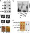
The Tsa1 and Tsa2 Prxs protect against Sup35 prion formation. (A) Subcellular distribution of Sup35 in WT (W303), tsa1, tsa2, and tsa1 tsa2 mutant cells grown to exponential phase in minimal SD media. Subcellular fractionation analysis of Sup35 was performed as described in Experimental Procedures using an anti-Sup35 polyclonal antibody. (T, total crude extract; S, soluble fraction; P, pellet fraction.) Representative gels are shown from at least three independent repeats. (B) Fluorescence micrographs of the W303 WT and tsa1 tsa2 mutant and [PSI+] and [psi] derivatives of 74D-694 carrying plasmid p6442 (CUP1-SUP35NM-GFP). (C) Detection of SDS-resistant Sup35 aggregates by SDD-AGE in cell lysates from WT (W303) tsa1, tsa2, and tsa1 tsa2 mutants, and [psi] [pin], [psi] [PIN+], and [PSI+] [PIN+] derivatives of 74D-694. The tsa1 tsa2 mutant was also grown in the presence of 3 mM GdnHCl. (D) WT (74D-694) and tsa1 tsa2 mutants were streaked on YEPD media and colony color visualized after 3 d growth. (E) [PSI+] prion formation was assayed in the WT (74D-694) and mutant strains by pink/white colony formation and growth on minimal media in the absence of adenine.
Fig. 3.
Fig. 3.
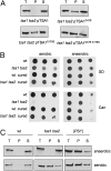
Prion formation in the tsa1 tsa2 mutant requires oxygen. (A) Subcellular distribution of Sup35 in the tsa1 tsa2 mutant cells containing WT Tsa1 (pTSA1) or cysteine mutants of Tsa1 (pTSA1C47S, pTSA1C170S, pTSA1C47S, C170S) as the sole source of TSA1. (B) [PSI+] prion formation assayed in cured [psi] [pin] and noncured WT (W303) [psi] [PIN+] and tsa1 tsa2 [PSI+] [PIN+] mutant strains assayed by growth on canavanine. The can1-100 allele in W303 contains a UAA nonsense mutation and translation termination at this stop codon confers resistance to canavanine. Cells were grown to stationary phase, diluted to A600s of 1.0, 0.1, and 0.01, and spotted onto minimal SD plates containing 80 μg/mL canavanine under aerobic or anaerobic conditions. (C) Subcellular distribution of Sup35 in the WT W303 [psi] [PIN+], tsa1 tsa2 [PSI+] [PIN+] mutant, and [PSI+] [PIN+] derivatives of 74D-694. Cells were grown under aerobic or anaerobic conditions in minimal SD media as described in the text.
Fig. 4.
Fig. 4.
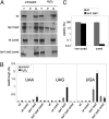
[PSI+] prion formation is induced by oxidative stress. (A) Subcellular distribution of Sup35 in cured and noncured WT (W303) and tsa1 tsa2 mutants grown in minimal SD media and treated with 100 μM H2O2 for 20 h. The levels of termination codon readthrough (B) and cell viability (C) were measured for the same strains and growth conditions as described in A. Percentage survival is expressed relative to untreated cultures.

References

    1. Wood ZA, Schröder E, Robin Harris J, Poole LB. Structure, mechanism and regulation of peroxiredoxins. Trends Biochem Sci. 2003;28:32–40. - PubMed
    1. Veal EA, Day AM, Morgan BA. Hydrogen peroxide sensing and signaling. Mol Cell. 2007;26:1–14. - PubMed
    1. Garrido EO, Grant CM. Role of thioredoxins in the response of Saccharomyces cerevisiae to oxidative stress induced by hydroperoxides. Mol Microbiol. 2002;43:993–1003. - PubMed
    1. Wong CM, Siu KL, Jin DY. Peroxiredoxin-null yeast cells are hypersensitive to oxidative stress and are genomically unstable. J Biol Chem. 2004;279:23207–23213. - PubMed
    1. Iraqui I, et al. Peroxiredoxin Tsa1 is the key peroxidase suppressing genome instability and protecting against cell death in Saccharomyces cerevisiae. PLoS Genet. 2009;5:e1000524. - PMC - PubMed

Publication types

MeSH terms