. 2010 Apr 6;107(14):6228-33.
doi: 10.1073/pnas.1001855107. Epub 2010 Mar 22.

Drosophila topo IIIalpha is required for the maintenance of mitochondrial genome and male germ-line stem cells

Affiliations

Drosophila topo IIIalpha is required for the maintenance of mitochondrial genome and male germ-line stem cells

Jianhong Wu et al. Proc Natl Acad Sci U S A. .

Abstract

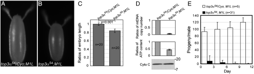
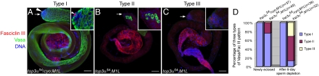
Topoisomerase IIIalpha (topo IIIalpha), a member of the conserved Type IA subfamily of topoisomerases, is required for the cell proliferation in mitotic tissues, but has a lesser effect on DNA endoreplication. The top3alpha gene encodes two forms of protein by utilizing alternative translation initiation sites: one (short form) with the nuclear localization signal only, exclusively localized in the nuclei, and the other (long form), retaining a mitochondrial import sequence at the N-terminus and the nuclear localization sequence at the C-terminus, localized primarily in the mitochondria, though with a small portion in the nuclei. Both forms of topo IIIalpha can rescue the viability of null mutants of top3alpha. No apparent defect is associated with the flies rescued by the long form; short-form-rescued flies (referred to as M1L), however, exhibit defects in fertilities. M1L females are sterile. They can lay eggs but with mitochondrial DNA (mtDNA) copy number and ATP content decreased by 20- and 2- to 3-fold, respectively, and they fail to hatch. Of the newly eclosed M1L males, 33% are completely sterile, whereas the rest have residual fertilities that are quickly lost in 6 days. The fertility loss of M1L males is caused by the disruption of the individualization complex and a progressive loss of germ-line stem cells. This study implicates topo IIIalpha in the maintenance of mtDNA and male germ-line stem cells, and thus is a causative candidate for genetic disorders associated with mtDNA depletion.


Conflict of interest statement

The authors declare no conflict of interest.

Figures

Fig. 1.
Fig. 1.
The topo IIIα is localized in both mitochondria and nuclei during spermatogenesis. (A) The apical tip of an adult testis. (B) A cyst of 16 spermacytes in the metaphase meiosis I, which is featured by the highly condensed chromatin and clustered mitochondria surrounding the equator spindle. (C) A cyst of onion stage spermatids. (D) Elongating spermatids at comet stage. (E) Elongated spermatids. AE are the merged double staining of topo IIIα (AE), and DNA (A′′E′′). (F) Merged triple staining of a cyst of onion stage spermatids with staining for topo IIIα, DNA, and cytochrome c (F, F′′, and F′′′, respectively). Abundant topo IIIα staining is colocalized with a mitochondrial marker, cytochrome c. Scale bars: 25 μm.
Fig. 2.
Fig. 2.
The unconserved amino (N) termini of topo IIIα contain mitochondrial import signals. (A) N-terminal sequences of topo IIIα of human, mouse, chicken, frog, and fruit fly. The mitochondrial import probabilities were obtained by using the algorithm developed by Claros and Vincens (21). Arrowhead indicates the mitochondrial endopeptidase cleavage sites (19). The second methionines are colored green. (B) The N-termini of proteins encoded by the wildtype and mutant transgenes of Drosophila. Methionines are colored green and the mutated amino acid is marked red. For M1L-YFP transgene, the first AUG was mutated to UUG, therefore the translation would start exclusively from the second AUG. (C) Confocal images of live S2 cells transfected with the plasmid DNA containing a transgene of topo IIIα-YFP, M1L-YFP, or M26L-YFP. Arrow indicates an S2 cell not transfected with transgenic vector. Scale bar, 10 μm. (D) Confocal images of fixed ovarioles of M1L-YFP and M26L-YFP transgenic females. Scale bar, 25 μm. (E) Subcellular fractionation and immunoblots. Mitochondria fractions (Lanes 1–3) and nuclei fractions (Lanes 4–6) were separated as described in Materials and Methods, and were subjected to SDS-PAGE, followed by Western blotting with antibodies against topo IIIα, lamin (nuclear marker) and cytochrome c (mitochondrial marker). Asterisk indicates the endogenous topo IIIα and arrow marks YFP fusion products.
Fig. 3.
Fig. 3.
The top3α gene and its null mutant top3α6 and top3α54. (A) Genomic structure of top3α gene and its mutants top3α6 and top3α54. Open triangles represent the remnant inserts derived from P-element and the dashed lines indicate the deleted sequences. Introns are shown as gray bars. (B) Mutants top3α6 and top3α54 were verified as null mutants by Western blotting. Lysate of ovaries was subjected to SDS-PAGE and the transferred protein bands were probed with antibodies against topo IIIα, and actin (loading control). Upper bands correspond to topo IIIα-YFP fusion protein, encoded by the transgene top3α-YFP, and the lower bands to the endogenous topo IIIα. (C, D) Brains and attached imaginal discs (Arrowheads) were dissected from wandering larvae of control (C) and top3α54 mutant (D) visualized with DAPI staining. Salivary gland nuclei (E, F) and fat body nuclei (G, H) of wandering larvae from control (E, G) and top3α54 mutants (F, H) were stained with DAPI.
Fig. 4.
Fig. 4.
The topo IIIα is required for mtDNA genome maintenance and fertilities of flies. (A, B) Eggs laid by M1L females, which are shorter than their heterozygous siblings, cannot hatch. (C) Embryo length of 20 eggs for each genotype was measured with Qcapture Pro version 5.1 (Qimaging) and analyzed with Student’s t test. (D) 0–1 hr embryos laid by M1L females have lower mtDNA copy number and ATP content, although cytochrome c level is not affected. (E) Residual fertility of M1L males is lost after 6 day sperm depletion.
Fig. 5.
Fig. 5.
The functions of topo IIIα in mitochondria are required for male germ-line stem cell maintenance. Testes dissected from the control (A) and M1L (B and C) males after 6 day sperm depletion (SI Materials and Methods) were fixed and stained with antibody against Vasa (marker for germ-line cells), Fasciclin III (marker for hub cells, the niche of germ-line stem cells), and DAPI. Insets (AC) highlight the tips, pointed by the arrows, of testes with higher magnification. Arrowhead in (A) indicates the hub cells marked by Fasciclin III. The Vasa/Fasciclin III immnunostaining patterns fell into three types: Type I (Panel A), with distinct Fasciclin III and Vasa signals, Type II (Panel B), without apparent Fasciclin III, but with germ-line cells scattered throughout the testis tube and Type III (Panel C), no germ-line cells present. (D) Quantification of three types of Fasciclin III/Vasa pattern for testes of males newly eclosed and after 6 day sperm depletion. Scale bars, 50 μm.

Similar articles

Cited by

References

    1. Schoeffler AJ, Berger JM. DNA topoisomerases: Harnessing and constraining energy to govern chromosome topology. Q Rev Biophys. 2008;41(1):41–101. - PubMed
    1. Wang JC. Cellular roles of DNA topoisomerases: A molecular perspective. Nat Rev Mol Cell Biol. 2002;3(6):430–440. - PubMed
    1. Li W, Wang JC. Mammalian DNA topoisomerase IIIalpha is essential in early embryogenesis. Proc Natl Acad Sci USA. 1998;95(3):1010–1013. - PMC - PubMed
    1. Plank JL, Chu SH, Pohlhaus JR, Wilson-Sali T, Hsieh TS. Drosophila melanogaster topoisomerase IIIalpha preferentially relaxes a positively or negatively supercoiled bubble substrate and is essential during development. J Biol Chem. 2005;280(5):3564–3573. - PubMed
    1. Krogh BO, Shuman S. A poxvirus-like type IB topoisomerase family in bacteria. Proc Natl Acad Sci USA. 2002;99(4):1853–1858. - PMC - PubMed

Publication types

MeSH terms

Substances