

Neurons sense nanoscale roughness with nanometer sensitivity
- PMID: 20308580
- PMCID: PMC2851967
- DOI: 10.1073/pnas.0914456107
Neurons sense nanoscale roughness with nanometer sensitivity
Abstract
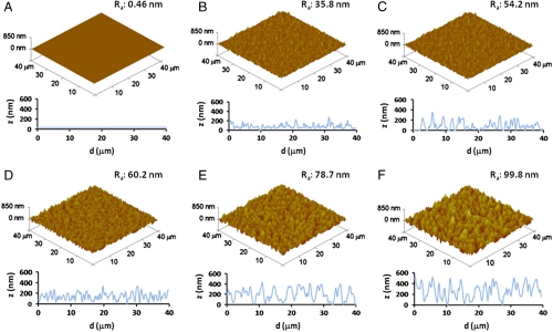
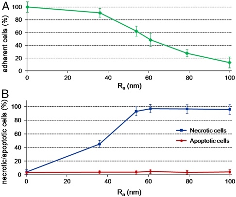
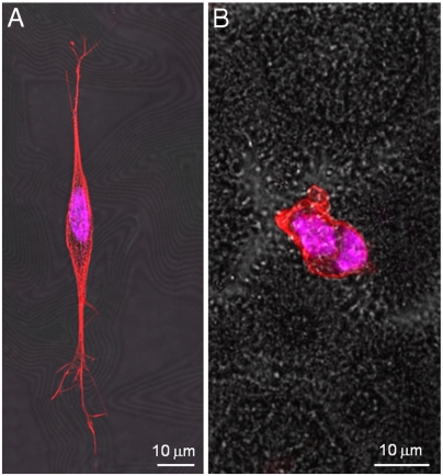
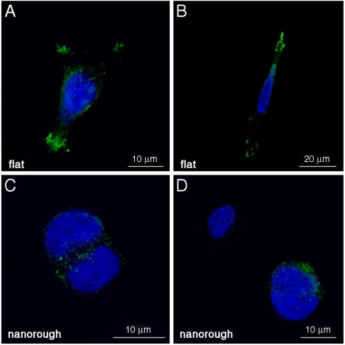
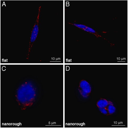
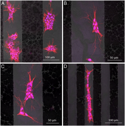
The interaction between cells and nanostructured materials is attracting increasing interest, because of the possibility to open up novel concepts for the design of smart nanobiomaterials with active biological functionalities. In this frame we investigated the response of human neuroblastoma cell line (SH-SY5Y) to gold surfaces with different levels of nanoroughness. To achieve a precise control of the nanoroughness with nanometer resolution, we exploited a wet chemistry approach based on spontaneous galvanic displacement reaction. We demonstrated that neurons sense and actively respond to the surface nanotopography, with a surprising sensitivity to variations of few nanometers. We showed that focal adhesion complexes, which allow cellular sensing, are strongly affected by nanostructured surfaces, leading to a marked decrease in cell adhesion. Moreover, cells adherent on nanorough surfaces exhibit loss of neuron polarity, Golgi apparatus fragmentation, nuclear condensation, and actin cytoskeleton that is not functionally organized. Apoptosis/necrosis assays established that nanoscale features induce cell death by necrosis, with a trend directly related to roughness values. Finally, by seeding SH-SY5Y cells onto micropatterned flat and nanorough gold surfaces, we demonstrated the possibility to realize substrates with cytophilic or cytophobic behavior, simply by fine-tuning their surface topography at nanometer scale. Specific and functional adhesion of cells occurred only onto flat gold stripes, with a clear self-alignment of neurons, delivering a simple and elegant approach for the design and development of biomaterials with precise nanostructure-triggered biological responses.
Conflict of interest statement
The authors declare no conflict of interest.
Figures
References
-
- Chen CS, Mrksich M, Huang S, Whitesides GM, Ingber DE. Geometric control of cell life and death. Science. 1997;276:1425–1428. - PubMed
-
- Geiger B, Spatz JP, Bershadsky AD. Environmental sensing through focal adhesions. Nat Rev Mol Cell Biol. 2009;10:21–33. - PubMed
-
- Spatz JP, Geiger B. Molecular engineering of cellular environments: Cell adhesion to nano-digital surfaces. Methods Cell Biol. 2007;83:89–111. - PubMed
MeSH terms
LinkOut - more resources
Full Text Sources
Other Literature Sources
