. 2010 Apr 6;107(14):6298-303.
doi: 10.1073/pnas.0914604107. Epub 2010 Mar 22.

Subunit interactions in bovine papillomavirus

Affiliations

Subunit interactions in bovine papillomavirus

Matthias Wolf et al. Proc Natl Acad Sci U S A. .

Abstract

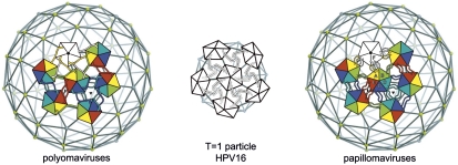
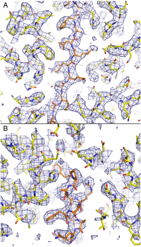
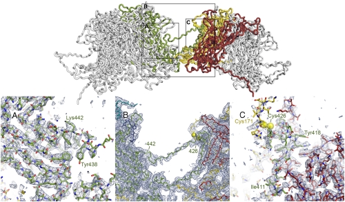
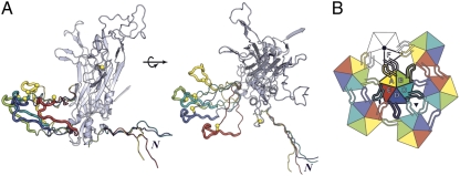
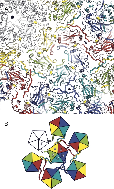
Papillomaviruses, members of a group of dsDNA viruses associated with epithelial growths and tumors, have compact capsids assembled from 72 pentamers of the protein L1. We have determined the structure of bovine papillomavirus by electron cryomicrosopy (cryoEM), at approximately 3.6 A resolution. The density map, obtained from single-particle analysis of approximately 4,000 particle images, shows the trace of the L1 polypeptide chain and reveals how the N- and C-terminal "arms" of a subunit (extensions from its beta-jelly-roll core) associate with a neighboring pentamer. Critical contacts come from the C-terminal arm, which loops out from the core of the subunit, forms contacts (including a disulfide) with two subunits in a neighboring pentamer, and reinserts into the pentamer from which it emanates. This trace corrects one feature of an earlier model. We discuss implications of the structure for virion assembly and for pathways of infectious viral entry. We suggest that it should be possible to obtain image reconstructions of comparable resolution from cryoEM images of asymmetric particles. From the work on papillomavirus described here, we estimate that such a reconstruction will require about 1.5 million images to achieve the same number of averaged asymmetric units; structural variability will increase this number substantially.


Conflict of interest statement

The authors declare no conflict of interest.

Figures

Fig. 1.
Fig. 1.
Diagrams of pentamer packing and C-terminal arm interchange in polyoma and papillomaviruses. In the full-size capsids of viruses from both groups, 72 pentamers of the major capsid protein (called VP1 in the polyomaviruses and L1 in the papillomaviruses) are arrayed at the vertices of a T = 7 d icosahedral lattice. Twelve pentamers (white) are centered on 5-fold symmetry axes, whereas the 60 others (6 of which are shown, colored) are at 6-coordinated positions. In a 12-pentamer (T = 1) icosahedral particle, obtained with N-terminally truncated L1 from HPV16, all 12 pentamers are on icosahedral 5-fold symmetry axes. Left: In the polyomaviruses, the VP1 C-terminal arm “invades” a neighboring pentamer, in the pattern shown, and “inserts” into its structure. Right: In the papillomaviruses, the L1 C-terminal arm invades a neighboring pentamer but turns around and inserts back into its pentamer of origin. The T = 1 shell has arm–arm interactions distinct from those in the virion.
Fig. 2.
Fig. 2.
Portions of the BPV cryoEM density map, in the 6-fold NCS-averaged region, with the refined molecular model superposed. The map is contoured at 3.2σ above the average density within the L1 β-barrel core. Side-chain densities in an α-helix (A) and a β-strand (B) lead to clear assignment of rotamers in many cases.
Fig. 3.
Fig. 3.
The L1 C-terminal arm. Top: The interface between two 6-coordinated pentamers. Polypeptide chains are in “worm” representation. The arm of the “green” subunit in the left-hand pentamer (the other subunits are in gray, for clarity) invades the right-hand pentamer, where it augments a β-sheet in the red subunit, forms a disulfide bond (yellow spheres) with the yellow subunit, and extends back to reinsert into its pentamer of origin. The N-to-C direction of the green arm is left-to-right at the “lower” part of the interface shown and right-to-left in the contact-free segment that retraverses the interpentamer gap. The outlined rectangles indicate regions shown in the lower part of the figure. Bottom: Details of the density map to show unambiguous tracing of the L1 arm. In (A) and (C), the 6-fold NCS-averaged map is contoured at 3.0σ above mean density. In (B), a region in which the NCS symmetry does not hold, the map is the final icosahedrally symmetrized reconstruction, contoured at 2.5σ. (A) Reinsertion of the arm into the left-hand pentamer. The PYAGFKF sequence (residues 437–443) where the arm reinserts can be assigned unambiguously, as can residues following it. (B) Connection between the disulfide (Cys426) and the PYAGF segment in (A). The sequence in this bridge is proline-rich in most papillomaviruses. (C) Invasion of the red subunit of the target pentamer (augmentation of the BIDG sheet) and disulfide bond between Cys426 in the arm and Cys171 in the yellow subunit. Assignment of sequence to the augmenting (green) strand [LxDxYR(Y/F) motif, from 412–418] is unambiguous. Cys171 is in a surface loop, less rigid than the core β-jelly roll, and the density of the yellow chain in the vicinity of Cys171 is correspondingly weaker than the core density of the red subunit.
Fig. 4.
Fig. 4.
Conformations of the BPV L1 arm. (A) Subunits A–E, superposed so that their core β-jelly rolls coincide (only one is shown, in gray), with N-terminal arms and extended part of C-terminal arms in color. The core of the subunit is in standard ribbon representation; the arms are in worm representation, in colors corresponding to those in the diagram in B and in Figs. 1,3, and 5. The gray arm, which has the same conformation as the yellow arm, is not shown. The parts of the C-terminal arms that contact subunits of the neighboring pentamer are shown as thicker worms; the conformation of these segments is the same for all subunits, because it is determined by conserved interactions (including the disulfide) with the target subunits. Yellow balls: The cysteines of the interpentamer disulfide. The N-terminal arms all have the same conformation C-terminal to residue 14 (the position at which the superposed arms splay apart). The two parts of A are related by a 90° rotation, as shown. (B) The icosahedral asymmetric unit, viewed from outside the particle, showing the pattern of C-terminal arm contacts.
Fig. 5.
Fig. 5.
View of BPV capsid from inside, looking outwards, centered along the axis of a 6-coordinated pentamer. (A) Ribbon representation, with N-terminal arms in worm representation, to distinguish them. Note the augmentation of a β-sheet in each subunit by the C-terminal segment of the neighboring subunit in the pentamer. (B) Icosahedral asymmetric unit, viewed from within the capsid. The pattern of N-terminal arm contacts is illustrated by the worm-like projections from each subunit. Note that N-terminal arms are ordered only on subunits A, C, D, and E.

References

    1. Howley PM, Lowy DR. Papillomaviridae. In: Knipe DM, Howley PM, editors. Fields Virology. Philadelphia: Lippincott Williams and Wilkins; 2007. pp. 2299–2354.
    1. Shi L, et al. GARDASIL: Prophylactic human papillomavirus vaccine development—From bench top to bed-side. Clin Pharmacol Ther. 2007;81:259–264. - PubMed
    1. Baker TS, et al. Structures of bovine and human papillomaviruses. Analysis by cryoelectron microscopy and three-dimensional image reconstruction. Biophys J. 1991;60:1445–1456. - PMC - PubMed
    1. Rayment I, Baker TS, Caspar DL, Murakami WT. Polyoma virus capsid structure at 22.5 A resolution. Nature. 1982;295:110–115. - PMC - PubMed
    1. Liddington RC, et al. Structure of simian virus 40 at 3.8-A resolution. Nature. 1991;354:278–284. - PubMed

Publication types

MeSH terms

LinkOut - more resources