. 2010 Apr 6;107(14):6258-63.
doi: 10.1073/pnas.1001062107. Epub 2010 Mar 22.

Lethal protein produced in response to competition between sibling bacterial colonies

Affiliations

Lethal protein produced in response to competition between sibling bacterial colonies

Avraham Be'er et al. Proc Natl Acad Sci U S A. .

Abstract

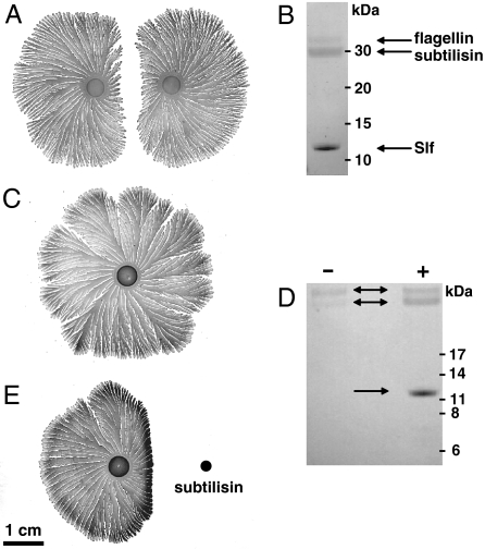
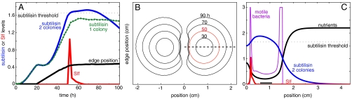
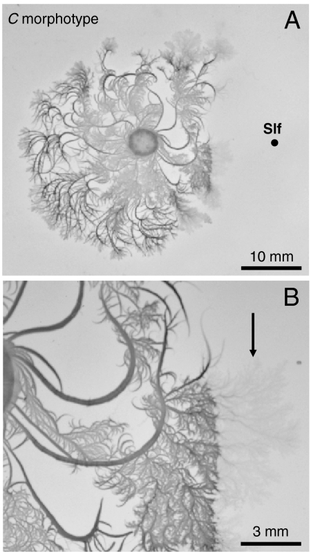
Sibling Paenibacillus dendritiformis bacterial colonies grown on low-nutrient agar medium mutually inhibit growth through secretion of a lethal factor. Analysis of secretions reveals the presence of subtilisin (a protease) and a 12 kDa protein, termed sibling lethal factor (Slf). Purified subtilisin promotes the growth and expansion of P. dendritiformis colonies, whereas Slf is lethal and lyses P. dendritiformis cells in culture. Slf is encoded by a gene belonging to a large family of bacterial genes of unknown function, and the gene is predicted to encode a protein of approximately 20 kDa, termed dendritiformis sibling bacteriocin. The 20 kDa recombinant protein was produced and found to be inactive, but exposure to subtilisin resulted in cleavage to the active, 12 kDa form. The experimental results, combined with mathematical modeling, show that subtilisin serves to regulate growth of the colony. Below a threshold concentration, subtilisin promotes colony growth and expansion. However, once it exceeds a threshold, as occurs at the interface between competing colonies, Slf is then secreted into the medium to rapidly reduce cell density by lysis of the bacterial cells. The presence of genes encoding homologs of dendritiformis sibling bacteriocin in other bacterial species suggests that this mechanism for self-regulation of colony growth might not be limited to P. dendritiformis.


Conflict of interest statement

The authors declare no conflict of interest.

Figures

Fig. 1.
Fig. 1.
Colonies of P. dendritiformis (T morphotype) grown for one week on 1.5% agar with 2 g/L peptone, and analysis of proteins released into the medium. (A) Two sibling colonies grown after simultaneous inoculation of an agar plate at the same distance from the plate’s center. (B) Proteins extracted from the agar between two colonies, analyzed by SDS–PAGE gel electrophoresis and identified by peptide sequencing as flagellin at 32 kDa, subtilisin at 30 kDa, and Slf at 12 kDa (Arrows). (C) A single colony of P. dendritiformis grown on an agar plate. (D) SDS–PAGE electrophoresis of proteins extracted from agar surrounding a single colony, without (indicated by - or with +) added subtilisin, as shown in panels C and E. (E) A single colony with subtilisin (0.1 mg dissolved in 5 μL) added 1 d after inoculation (Black Dot).
Fig. 2.
Fig. 2.
The list of the 173 amino acids of the DfsB protein corresponding to the dfsB gene. Large letters indicate the segment in position 5-169 amino acids, which is associated with a conserved Pfam family domain. Bold letters indicate the segment of the isolated protein Slf. The detected peptide (Edman sequencing) is underlined.
Fig. 3.
Fig. 3.
Effect of subtilisin and Slf on colony growth. (A) Subtilisin was added (at the positions indicated by arrows) to single colonies growing on an agar gel, 1 d after inoculation; the images give the time elapsed (in hours) after subtilisin was added. The amount of subtilisin dissolved in a 5 μL water inoculation droplet was: Upper row, 0.001 mg; only attraction of growing colonies was observed. Middle row, 0.01 mg; attraction occurred only initially. Lower row, 0.1 mg; only inhibition was observed (see Fig. 1E). (B) Subtilisin (Carlsberg, P8038 Sigma) was added in different amounts (20, 10, 5, 2.5, 0.5, 0.25, 0.05, 0.025, 0.005, 0.0025, 0.0005, 0.00025, 0.00005 mg) to low-nutrient liquid media at 30 °C, after each tube was inoculated with 1 × 106 bacteria taken from a rich liquid media culture. The optical density of each tube was measured 24 h after inoculation (OD = 1 at 650 nm corresponds to approximately 108 bacteria/mL, verified by counting colonies on LB plates after appropriate dilution). The arrow marks the amount of subtilisin (0.1 mg) added to low-nutrient media agar plates near a growing colony to show strong inhibition and cell death. Error bars stem from 5 independent experiments. (C) Slf was introduced at the black dot near a single growing colony 4 days after inoculation. The faint region (shown at higher magnification in the inset) corresponds to lysed cells; growing branches were absent. Only a single band appeared in the gel electrophoresis results for the isolated Slf (Insert, Lower Right). In both A and C the growth conditions were 1.5% agar with 2 g/L peptone nutrient.
Fig. 4.
Fig. 4.
Numerical simulation of colony competition. (A) The subtilisin levels (in arbitrary units) at the moving edge of a colony as a function of time, both for a single colony (Green Dots) and for the inhibited interface in the case of two colonies (Blue Line). After 40 h, a competing colony senses its neighbor, and after 50 h the subtilisin level crosses a prefixed threshold (Horizontal Dashed Black Line). The red curve shows the level of Slf for the inhibited colony; no Slf is secreted in the single colony case. The black line shows the position of the edge of a colony growing toward a neighbor. The colony’s edge starts to move after a lag time of 20 h, and then begins slowing down just before Slf is secreted. (B) The positions of the edges of competing colonies at 30, 50, 70, and 90 h. (C) Levels of motile bacteria (Purple), subtilisin (Blue), nutrients (Black), and Slf (Red), measured along the horizontal dashed line in B, 50 h after inoculation. The gray rectangle represents the initial inoculation droplet. For each panel the model parameters and the initial conditions were the same; the initial distance between the colonies was 1.2 cm and the diameter of the dish was 8.8 cm, as in the experiments.
Fig. 5.
Fig. 5.
Slf (Black Point) placed near a single growing P. dendritiformis C morphotype colony (0.7% agar gel with 2 g/L peptone nutrient). (A) Inhibition of the colony. (B) Magnified image, which has a faint region (marked with an arrow) where bacteria are lysed. The concentration of Slf is same as in Fig. 3C.

References

    1. Errington J. Regulation of endospore formation in Bacillus subtilis. Nat Rev Microbiol. 2003;1:117–126. - PubMed
    1. Gonzalez-Pastor JE, Hobbs EC, Losick R. Cannibalism by sporulating bacteria. Science. 2003;301:510–513. - PubMed
    1. Ellermeier CD, Hobbs EC, Gonzalez-Pastor JE, Losick R. A three-protein signaling pathway governing immunity to a bacterial cannibalism toxin. Cell. 2006;124:549–559. - PubMed
    1. Claverys JP, Havarstein LS. Cannibalism and fratricide: mechanisms and raisons d’etre. Nature. 2007;5:219–229. - PubMed
    1. Guiral S, Mitchell TJ, Martin B, Claverys JP. Competence-programmed predation of noncompetent cells in the human pathogen Streptococcus pneumoniae: Genetic requirements. Proc Natl Acad Sci USA. 2005;102:8710–8715. - PMC - PubMed

Publication types

MeSH terms

Associated data

LinkOut - more resources