. 2009 Aug 20;2(4):237-44.
doi: 10.1007/s12308-009-0043-y.

Rare lymphoid malignancies of the breast: a report of two cases illustrating potential diagnostic pitfalls

Rare lymphoid malignancies of the breast: a report of two cases illustrating potential diagnostic pitfalls

Evan A Farkash et al. J Hematop. .

Abstract

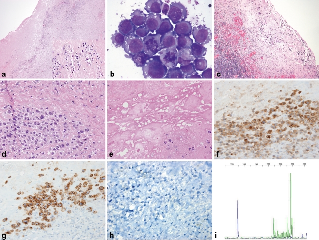
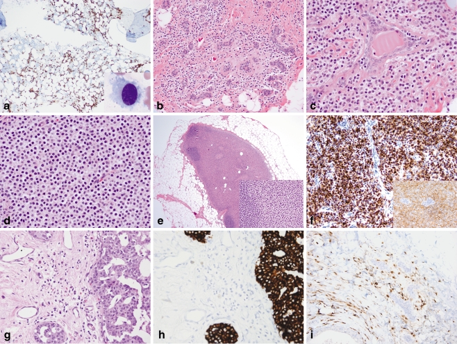
Breast involvement by lymphoma is uncommon and poses challenges in diagnosis. Lymphomas may clinically, radiologically, and morphologically mimic both benign and neoplastic conditions. We describe two cases of lymphoid malignancies predominantly involving the breast, both presenting diagnostic dilemmas. The first case, ALK-negative anaplastic large-cell lymphoma involving a seroma associated with a breast implant, is an emerging clinicopathologic entity. Anaplastic large-cell lymphoma has been identified in association with breast implants and seroma formation relatively recently. The second case, hairy cell leukemia involving the breast and ipsilateral axillary sentinel lymph node, is, to our knowledge, the first reported case of hairy cell leukemia involving the breast at the time of diagnosis. While a localized bone lesion was present at time of diagnosis, bone marrow involvement was relatively mild in comparison to that seen in the breast and lymph node. In the first case, lymphoma occurred in a clinical setting where malignancy was unsuspected, highlighting the importance of careful morphologic evaluation of paucicellular samples, as well as awareness of rare clinicopathologic entities, in avoiding a misdiagnosis of a benign inflammatory infiltrate. In the second case, the lymphoid neoplasm exhibited classic morphologic and immunophenotypic features, but presented at an unusual site of involvement. Knowledge of the patient's concurrent diagnosis of hairy cell leukemia involving the bone marrow and bone helped avoid a misdiagnosis of carcinoma rather than lymphoma.

Keywords: Anaplastic large cell lymphoma; Anaplastic lymphoma kinase; Breast; Breast implant; Hairy cell leukemia; Primary breast lymphoma; Seroma; T-cell neoplasm.


Figures

Fig. 1
Fig. 1
ALK-negative ALCL arising in a breast peri-implant capsule and associated seroma. a Low-power examination of the initial implant capsulectomy specimen reveals an apparent benign chronic inflammatory infiltrate in a background of dense fibrosis (H&E, ×40); however, atypical, but focally necrotic, large cells with hyperchromatic, irregular nuclei, and abundant eosinophilic cytoplasm are visible on high magnification (inset, H&E, ×400). b Large malignant-appearing cells with high nuclear-to-cytoplasmic ratios, prominent nucleoli, vacuolated cytoplasm, and mitotic figures are present in a cytospin of the seroma aspirate (Wright-Giemsa stain, ×1,000). c The second implant capsulectomy contains fibrosis, chronic inflammation, and more conspicuous clusters of large, atypical cells adjacent to the seroma cavity (H&E, ×100). d Viable clusters of large infiltrating cells have pleomorphic nuclei and prominent nucleoli with occasional “hallmark cells” present (H&E, ×400). e The implant capsule contains focal deposits of refringent, nonpolarizable material consistent with silicone adjacent to partially viable clusters of large atypical cells (H&E, ×400). fh Immunohistochemical staining demonstrates the neoplastic cells to be positive for CD4 (f) and CD30 (g), but negative for ALK-1 (h; ×400). i T-cell receptor PCR identified a clonal rearrangement with Vγ10 primers (174.5 bp, blue tracing), as well as an indeterminate peak with Vγ1-8 primers (218.6 bp, green tracing), consistent with the presence of a clonal T-cell population
Fig. 2
Fig. 2
Hairy cell leukemia involving the bone marrow, breast, and axillary sentinel lymph node. a A posterior iliac crest bone marrow aspirate smear contains medium-sized lymphoid cells with oval nuclei, dispersed chromatin, and abundant pale cytoplasm with circumferential cytoplasmic projections (inset, Wright-Giemsa, ×1,000). CD20 stain of the core biopsy highlights the interstitial pattern of marrow involvement characteristic of hairy cell leukemia (×100). bd Left breast lumpectomy containing prominent monomorphic infiltrates of small discohesive cells with abundant pale cytoplasm and round to oval nuclei present singly and in linear strands surrounding benign lobules (b; H&E, ×200), as larger clusters and aggregates surrounded by thin bands of fibrous stroma (c; H&E, ×400), as well as in solid sheets (d; H&E, ×400). DCIS was present elsewhere (not shown). e The sentinel lymph node shows massive replacement of the paracortex and hilum by cells with abundant pale eosinophilic cytoplasm and oval to spindled nuclei, morphologically similar to those in the breast (inset, H&E, ×400); residual primary follicles are present at the periphery of the node (H&E, ×20). f Immunohistochemistry of the breast specimen reveals infiltrating cells to be CD20-positive (inset) and strongly DBA.44-positive (×400). g Subsequent mastectomy demonstrates an infiltrate of monomorphic cells present singly and in loose linear arrays adjacent to residual cribriform DCIS (H&E, ×400). hi A cytokeratin cocktail immunostain (h) of the mastectomy specimen highlights DCIS, but not the infiltrating cells, which are positive for CD20 (i; ×400)

References

    1. Lamovec J, Wotherspoon A, Jacquemier J. Malignant lymphoma and metastatic tumours. In: Tavassoli F, Devilee P, editors. Tumors of the breast and female genital organs. World Health Organization classification of tumours. Lyon: IARC; 2003. pp. 107–109.
    1. Brogi E, Harris NL. Lymphomas of the breast: pathology and clinical behavior. Semin Oncol. 1999;26:357–364. - PubMed
    1. Talwalkar SS, Miranda RN, Valbuena JR, Routbort MJ, Martin AW, Medeiros LJ. Lymphomas involving the breast: a study of 106 cases comparing localized and disseminated neoplasms. Am J Surg Pathol. 2008;32:1299–1309. doi: 10.1097/PAS.0b013e318165eb50. - DOI - PubMed
    1. Kuper-Hommel MJ, Snijder S, Janssen-Heijnen ML, et al. Treatment and survival of 38 female breast lymphomas: a population-based study with clinical and pathological reviews. Ann Hematol. 2003;82:397–404. doi: 10.1007/s00277-003-0664-7. - DOI - PubMed
    1. Wiseman C, Liao KT. Primary lymphoma of the breast. Cancer. 1972;29:1705–1712. doi: 10.1002/1097-0142(197206)29:6<1705::AID-CNCR2820290640>3.0.CO;2-I. - DOI - PubMed

LinkOut - more resources