. 2011 Jan 1;128(1):132-43.
doi: 10.1002/ijc.25330.

Prognostic significance of differentially expressed miRNAs in esophageal cancer

Affiliations

Prognostic significance of differentially expressed miRNAs in esophageal cancer

Yuxin Hu et al. Int J Cancer. .

Abstract

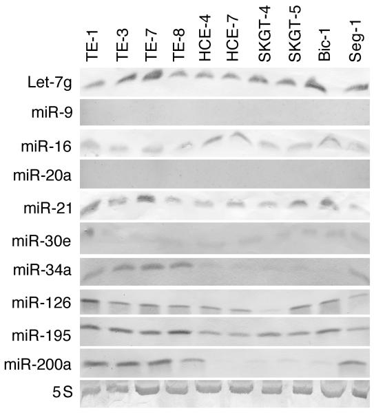
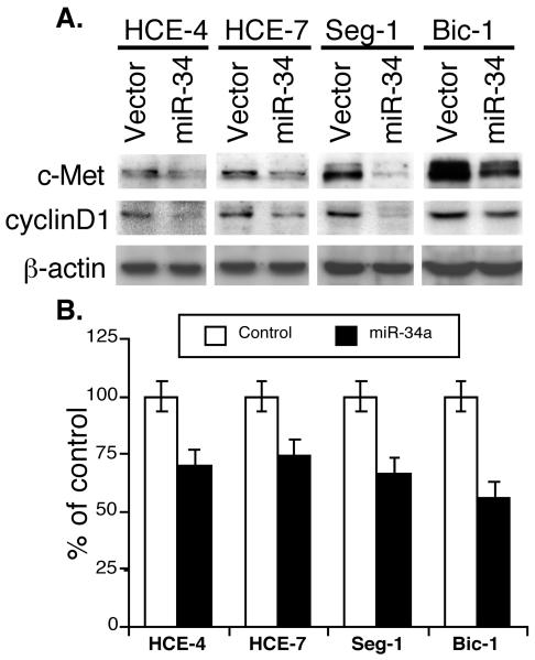
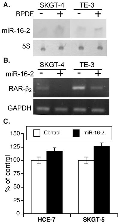
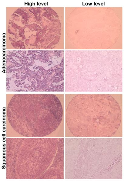
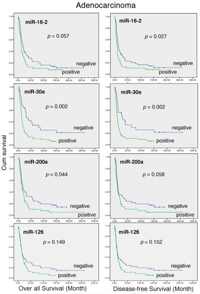
Altered microRNA (miRNA) expression has been found to promote carcinogenesis, but little is known about the role of miRNAs in esophageal cancer. In this study, we selected 10 miRNAs and analyzed their expression in 10 esophageal cancer cell lines and 158 tissue specimens using Northern blotting and in situ hybridization, respectively. We found that Let-7g, miR-21 and miR-195p were expressed in all 10 cell lines, miR-9 and miR-20a were not expressed in any of the cell lines, and miR-16-2, miR-30e, miR-34a, miR-126 and miR-200a were expressed in some of the cell lines but not others. In addition, transient transfection of miR-34a inhibited c-Met and cyclin D1 expression and esophageal cancer cell proliferation, whereas miR-16-2 suppressed RAR-β(2) expression and increased tumor cell proliferation. Furthermore, we found that miR-126 expression was associated with tumor cell dedifferentiation and lymph node metastasis, miR-16-2 was associated with lymph node metastasis, and miR-195p was associated with higher pathologic disease stages in patients with esophageal adenocarcinoma. Kaplan-Meier analysis showed that miR-16-2 expression and miR-30e expression were associated with shorter overall and disease-free survival in all esophageal cancer patients. In addition, miR-16-2, miR-30e and miR-200a expression were associated with shorter overall and disease-free survival in patients with esophageal adenocarcinoma; however, miR-16-2, miR-30e and miR-200a expression were not associated with overall or disease-free survival in squamous cell carcinoma patients. Our data indicate that further evaluation of miR-30e and miR-16-2 as prognostic biomarkers is warranted in patients with esophageal adenocarcinoma. In addition, the role of miR-34a in esophageal cancer also warrants further study.


Figures

Figure 1
Figure 1
Nonradioactive Northern blotting analysis of miRNA expression. Esophageal squamous cell carcinoma cell lines (TE-1, TE-3, TE-7, TE-8, HCE-4, and HCE-7) and adenocarcinoma cell lines (SKGT-4, SKGT-5, Seg-1 and Bic-1) were grown in Dulbecco's modified Eagle medium, and total RNA was extracted for Northern blot analysis with digoxigenin-labeled and locked nucleic acid–modified miRNA probes. The blots were then scanned and plotted.
Figure 2
Figure 2
miR-34a suppression of gene expression and proliferation of esophageal cancer cells. A, Western blotting. Esophageal squamous cell carcinoma lines HCE-4 and HCE-7 and adenocarcinoma cell lines Seg-1 and Bic-1 were grown in monolayer and transiently transfected with pRNA-u6.3/miR-34a vector or vector-only control and treated with G418 for 3 days. The total cellular protein was then extracted and subjected to Western blotting analysis of c-Met and cyclin D1 expression. B, Cell proliferation assay. Cells were grown in monolayer and transiently transfected with either pCMS/EGFP plus pRNA-u6.3/miR-34a vector or pCMS/EGFP plus pRNA-u6.3 vector. Cells were then fixed with 4% paraformaldehyde and permeabilized in Triton X-100. Afterward, cells were immunostained for Ki-67 expression, and approximately 200 cells were counted for positive GFP staining f (green) and positive or negative Ki-67 staining (red). The percentage of control of cell proliferation was then calculated.
Figure 3
Figure 3
The role of miR-16-2 in esophageal cancer cells. A, Northern blotting. Esophageal adenocarcinoma SKGT-4 cells and squamous cell carcinoma TE-3 cells were grown and treated with 1 μl BPDE for 12 h and RNA was isolated and subjected to northern blot analysis. B, SKGT-4 and TE-3 cells were grown in monolayer and transiently transfected with pRNA-u6.3/miR-16-2 vector or vector-only control and treated with G418 for 3 days. RNA from the cells was isolated and subjected to RT-PCR analysis of RAR-β2 expression. C, Esophageal adenocarcinoma SKGT-5 cells and squamous cell carcinoma HEC-7 cells were grown and transiently transfected with pRNA-u6.3/miR-16-2 vector or vector-only control and treated with G418 for 4 days. The cells were then subjected to cell viability assay.
Figure 4
Figure 4
Detection of miRNA expression in esophageal tissue microarrays using in situ hybridization. A tissue microarray contained 158 available cases of esophageal cancer, and three tissue spots for each case were on the same sections. A total of three sections were hybridized with each digoxigenin-labeled miRNA probe and then scored for expression of each miRNA.
Figure 5
Figure 5
Relationship between miRNA expression and survival in all esophageal cancer patients. The tissue microarrays were hybridized in situ with digoxigenin-labeled miRNA probes and then scored for high and low expression of each miRNA. The data on the high and low miRNA expression were then statistically analyzed against the overall and disease-free survival data of the 158 patients with esophageal cancer using the Kaplan-Meier method.
Figure 6
Figure 6
Relationship between miRNA expression and survival in the patients with esophageal adenocarcinoma. The tissue microarrays were hybridized in situ with digoxigenin-labeled miRNA probes and then scored for high and low expression of each miRNA. The data on the high and low miRNA expression were then statistically analyzed against the overall and disease-free survival data of the 99 patients with esophageal adenocarcinoma using the Kaplan-Meier method.

References

    1. Blot W. Esophageal cancer trends and risk factors. Semin Oncol. 1994;21:403–10. - PubMed
    1. van Soest EM, Dieleman JP, Siersema PD, Sturkenboom MC, Kuipers EJ. Increasing incidence of Barrett's oesophagus in the general population. Gut. 2005;54:1062–6. - PMC - PubMed
    1. Chen X, Yang CS. Esophageal adenocarcinoma: a review and perspectives on the mechanism of carcinogenesis and chemoprevention. Carcinogenesis. 2001;22:1119–29. - PubMed
    1. Reid BJ, Blount PL, Rabinovitch PS. Biomarkers in Barrett's esophagus. Gastrointest Endosc Clin N Am. 2003;13:369–97. - PubMed
    1. Spechler SJ. Barrett's esophagus: a molecular perspective. Curr Gastroenterol Rep. 2005;7:177–81. - PubMed

Publication types

MeSH terms