

Designer nanoparticles: incorporating size, shape and triggered release into nanoscale drug carriers
- PMID: 20331355
- PMCID: PMC2845970
- DOI: 10.1517/17425240903579971
Designer nanoparticles: incorporating size, shape and triggered release into nanoscale drug carriers
Abstract
Importance of the field: Although significant progress has been made in delivering therapeutic agents through micro and nanocarriers, precise control over in vivo biodistribution and disease-responsive drug release has been difficult to achieve. This is critical for the success of next generation drug delivery devices, as newer drugs, designed to interfere with cellular functions, must be efficiently and specifically delivered to diseased cells. The chief constraint in achieving this has been our limited repertoire of particle synthesis methods, especially at the nanoscale. Recent developments in generating shape-specific nanocarriers and the potential to combine stimuli-responsive release with nanoscale delivery devices show great promise in overcoming these limitations.
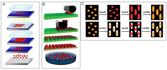
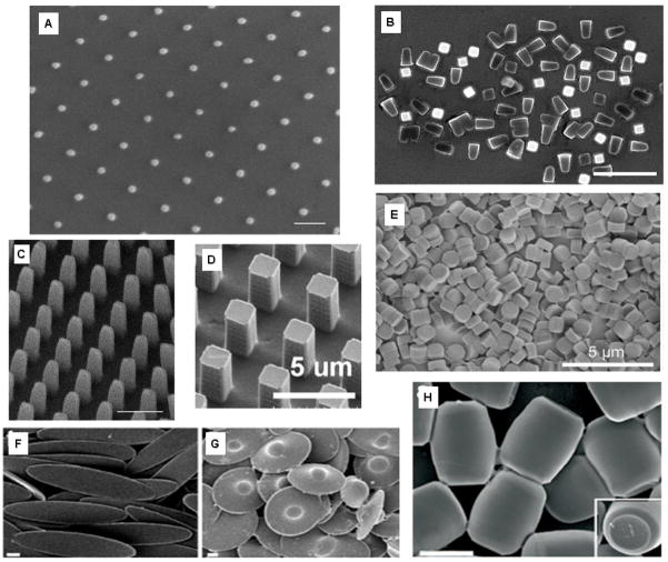
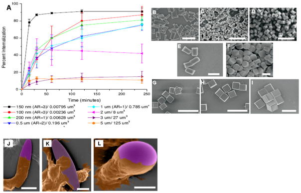
Areas covered in this review: How recent advances in fabrication technology allow synthesis of highly monodisperse, stimuli-responsive, drug-carrying nanoparticles of precise geometries is discussed. How particle properties, specifically shape and stimuli responsiveness, affect biodistribution, cellular uptake and drug release is also reviewed.
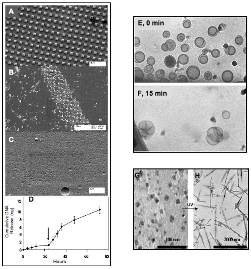
What the reader will gain: The reader is introduced to recent developments in intelligent drug nanocarriers and new nanofabrication approaches that can be combined with disease-responsive biomaterials. This will provide insight into the importance of controlling particle geometry and incorporating stimuli-responsive materials into drug delivery.
Take home message: The integration of responsive biomaterials into shape-specific nanocarriers is one of the most promising avenues towards the development of next generation, advanced drug delivery systems.
Conflict of interest statement
Figures
Similar articles
-
Particle shape effects in vitro and in vivo.Front Biosci (Schol Ed). 2012 Jun 1;4(4):1344-53. doi: 10.2741/s336. Front Biosci (Schol Ed). 2012. PMID: 22652876 Review.
-
Applications of stimuli-responsive nanoscale drug delivery systems in translational research.Drug Discov Today. 2018 May;23(5):1043-1052. doi: 10.1016/j.drudis.2017.11.009. Epub 2017 Nov 16. Drug Discov Today. 2018. PMID: 29155366 Review.
-
Disease-responsive drug delivery: the next generation of smart delivery devices.Curr Drug Metab. 2012 Jan;13(1):42-9. doi: 10.2174/138920012798356880. Curr Drug Metab. 2012. PMID: 22385534 Review.
-
Non-spherical micro- and nanoparticles: fabrication, characterization and drug delivery applications.Expert Opin Drug Deliv. 2015 Mar;12(3):481-92. doi: 10.1517/17425247.2015.963055. Epub 2014 Oct 18. Expert Opin Drug Deliv. 2015. PMID: 25327886 Review.
-
[Reactive oxygen species stimuli-responsive nanocarriers].Se Pu. 2021 Feb;39(2):118-124. doi: 10.3724/SP.J.1123.2020.11014. Se Pu. 2021. PMID: 34227343 Free PMC article. Review. Chinese.
Cited by
-
Functionalization of Cellulose Nanocrystals with PEG-Metal-Chelating Block Copolymers via Controlled Conjugation in Aqueous Media.ACS Omega. 2016 Jul 31;1(1):93-107. doi: 10.1021/acsomega.6b00055. Epub 2016 Jul 18. ACS Omega. 2016. PMID: 30023474 Free PMC article.
-
Critical parameters for design and development of multivalent nanoconstructs: recent trends.Drug Deliv Transl Res. 2022 Oct;12(10):2335-2358. doi: 10.1007/s13346-021-01103-4. Epub 2022 Jan 11. Drug Deliv Transl Res. 2022. PMID: 35013982 Free PMC article. Review.
-
Quality by design (QbD) approach in processing polymeric nanoparticles loading anticancer drugs by high pressure homogenizer.Heliyon. 2020 Apr 29;6(4):e03846. doi: 10.1016/j.heliyon.2020.e03846. eCollection 2020 Apr. Heliyon. 2020. PMID: 32373744 Free PMC article. Review.
-
Peeking into the future: Transdermal patches for the delivery of micronutrient supplements.Metabol Open. 2021 Jul 13;11:100109. doi: 10.1016/j.metop.2021.100109. eCollection 2021 Sep. Metabol Open. 2021. PMID: 34337377 Free PMC article.
-
Glycyrrhizic Acid Formulated in Hydrotalcite Nanocarriers Intended to Act as a Hepatoprotective Agent.AAPS J. 2024 Nov 19;27(1):2. doi: 10.1208/s12248-024-00986-8. AAPS J. 2024. PMID: 39562487
References
-
- Ferrari M. Cancer nanotechnology: opportunities and challenges. Nature Reviews Cancer. 2005;5(3):161–71. - PubMed
-
- Peer D, Karp JM, Hong S, et al. Nanocarriers as an emerging platform for cancer therapy. Nature Nanotechnology. 2007;2(12):751–60. - PubMed
-
- Zhang SF, Uludag H. Nanoparticulate Systems for Growth Factor Delivery. Pharmaceutical Research. 2009;26(7):1561–80. - PubMed
-
- Fenske DB, Chonn A, Cullis PR. Liposomal Nanomedicines: An Emerging Field. Toxicologic Pathology. 2008;36(1):21–9. - PubMed
-
- Duncan R. The dawning era of polymer therapeutics.(Report) Nature Reviews Drug Discovery. 2003;2(5):347. 14. - PubMed
Publication types
MeSH terms
Substances
Grants and funding
LinkOut - more resources
Full Text Sources
Other Literature Sources
Medical
Research Materials