. 2010 Jun;9(3):398-409.
doi: 10.1111/j.1474-9726.2010.00567.x. Epub 2010 Mar 13.

Ionizing radiation-induced long-term expression of senescence markers in mice is independent of p53 and immune status

Affiliations

Ionizing radiation-induced long-term expression of senescence markers in mice is independent of p53 and immune status

Oanh N L Le et al. Aging Cell. 2010 Jun.

Abstract

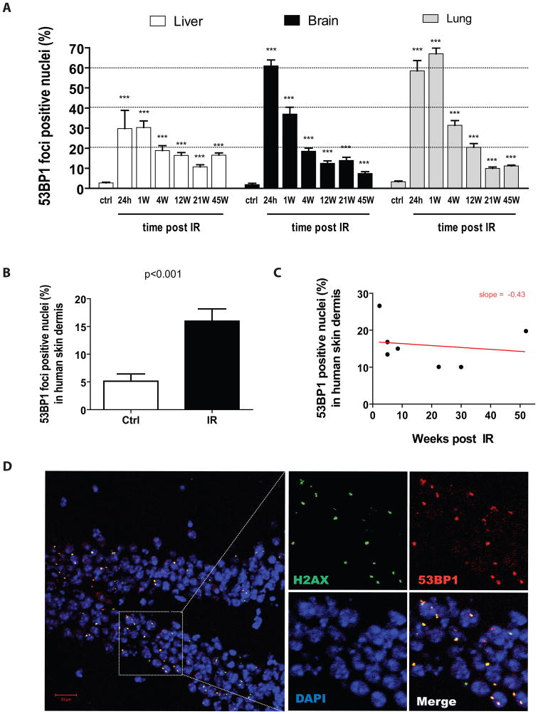
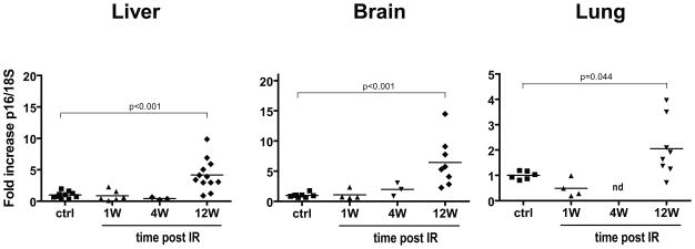
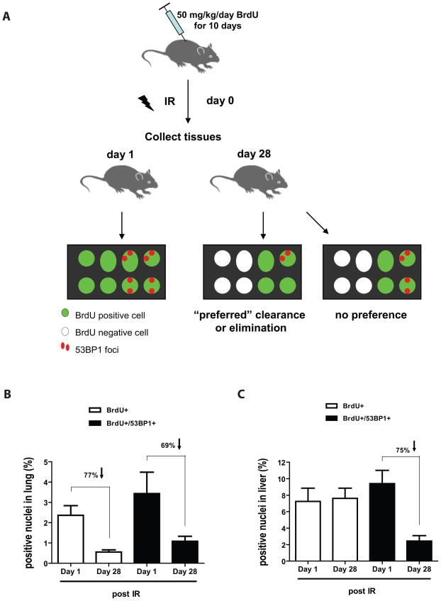
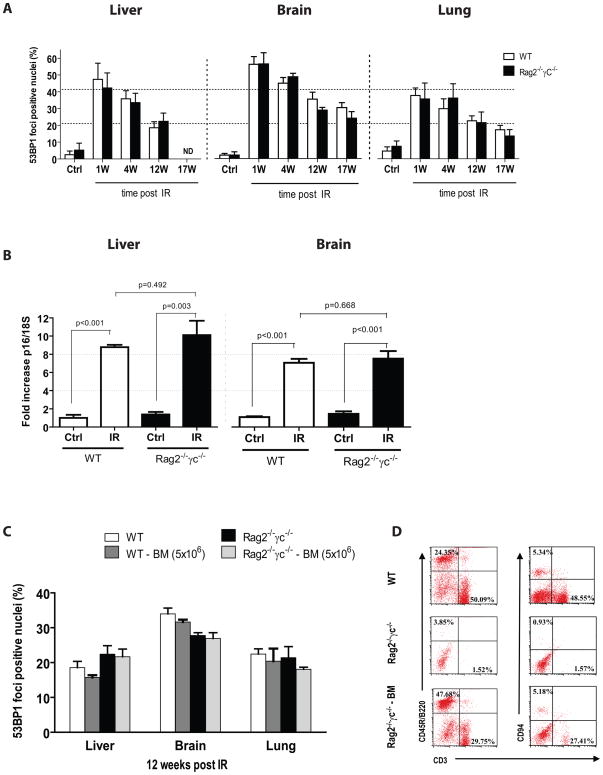
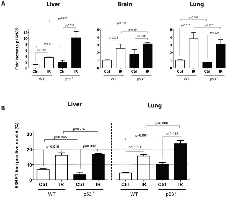
Exposure to IR has been shown to induce the formation of senescence markers, a phenotype that coincides with lifelong delayed repair and regeneration of irradiated tissues. We hypothesized that IR-induced senescence markers could persist long-term in vivo, possibly contributing to the permanent reduction in tissue functionality. Here, we show that mouse tissues exposed to a sublethal dose of IR display persistent (up to 45 weeks, the maximum time analyzed) DNA damage foci and increased p16(INK4a) expression, two hallmarks of cellular senescence and aging. BrdU-labeling experiments revealed that IR-induced damaged cells are preferentially eliminated, at least partially, in a tissue-dependent manner. Unexpectedly, the accumulation of damaged cells was found to occur independent from the DNA damage response modulator p53, and from an intact immune system, as their levels were similar in wild-type and Rag2(-/-) gammaC(-/-) mice, the latter being deficient in T, B, and NK cells. Together, our results provide compelling evidence that exposure to IR induces long-term expression of senescence markers in vivo, an effect that may contribute to the reduced tissue functionality observed in cancer survivors.


Conflict of interest statement

Conflict of interest: The authors have declared that no conflict of interest exists.

Figures

Figure 1
Figure 1. Exposure to IR induces persistent DNA damage foci-positive cells in mouse and human tissues
A) Fraction of cells containing 53BP1 DNA damage foci in liver, brain and lung tissues collected from control (ctrl) and from irradiated (8 Gy TBI) C57BL/6 mice sacrificed at the indicated time in weeks (W) post IR. Data are mean values ± s.e.m (n= 4–7). Statistical significance: *** P < 0.001 was obtained by performing a Student’s t-test relative to control. No increase in the fraction of DNA damage foci-positive cells was observed in control non-irradiated mice until they reached two years of age (see figure S1). B) Proportion of cells containing persistent 53BP1 DNA damage foci in the dermal region of skin collected from cancer patients who, as part of their treatments, received 12 Gy TBI (median of 8 weeks prior from being biopsied) and from age-matched control non-irradiated patients (n = 7–9 per group). p value was obtained by performing a Student’s t-test relative to control. C) Correlation of the amount of 53BP1 DNA damage foci-containing cells to the time to which the biopsies was obtained post exposure to IR. D) Detection by confocal microscopy of 53BP1 (red) and γ-H2AX (green) DNA damage foci from the hippocampus of a mouse sacrificed 12 weeks post its exposure to IR. Nuclei were stained with DAPI.
Figure 2
Figure 2. Exposure to IR induces delayed p16INK4a expression in mouse tissues
RNA was isolated from homogenized liver, brain and lung tissues collected from control (ctrl) and from irradiated (8 Gy TBI) C57BL/6 mice sacrificed at the indicated time in weeks (W) post IR. RNA was then used to determined p16INK4a expression by quantitative real-time PCR (n = 3–12, each symbol representing an individual mouse). p values were obtained by performing a Student’s t-test relative to control. nd = not determined.
Figure 3
Figure 3. Clearance of IR-induced DNA damaged cells is tissue dependent
A) Schematic representation of the protocol used and the expected outcomes of BrdU+ cells containing or not 53BP1 DNA damage foci. B and C) Proportion of cells labelled with BrdU (alone) or BrdU and 53BP1 foci at 1 or 28 days post exposure to 8 Gy TBI. Indicated is the proportion of cells that have lost BrdU signal during the 28 days period post exposure to IR. n = 4 in each group.
Figure 4
Figure 4. IR-induced persistent DNA damage foci and p16INK4a expression is independent of T, B and NK cell functions
A) Proportion of cells containing 53BP1 DNA damage foci in liver, brain and lung tissues collected from wild type (WT) or Rag2−/−γC−/− immune deficient mice sacrificed at the indicated time in weeks (W) post 6 Gy TBI (n = 3–5 mice per group). nd = not determined. No statistical difference was observed between groups of mice at all time point analyzed post IR. B) Expression of p16INK4a as determined by quantitative real-time PCR using RNA isolated from homogenized liver or brain tissues of wild type and Rag2−/−γC−/− mice 17–18 weeks post 6 Gy TBI, p values were obtained by performing a Student’s t-test. C) As described for panel A, proportion of cells containing 53BP1 DNA damage foci 12 weeks post IR in wild type and Rag2−/−γC−/− mice transplanted or not with 5×10⁶ bone marrow cells (BM) collected from a wild type donor mouse (n = 4 per group). No statistical difference was observed between transplanted and non-transplanted groups. D) Representative flow cytometry profiles showing the proportion of T cells (CD3+), B cells (CD45R/B220) and NK cells (CD94+, CD3) in blood collected from wild type (WT), and Rag2−/−γC−/− mice transplanted (Rag2−/−γC−/− BM) or not with bone marrow cells.
Figure 5
Figure 5. p53 is not required for the induction of persistent DNA damage foci and increased p16INK4a expression post IR
A) Expression of p16INK4a was determined by quantitative real-time PCR on liver, brain and lung tissues collected from age-match wild type (WT) or p53−/− deficient mice (n = 3–5 mice per group) 8 weeks following their exposure to 5 Gy TBI. B) Proportion of cells containing 53BP1 DNA damage foci in tissues collected from mice as described in panel A. p values were obtained by performing a Student’s t-test.

References

    1. Baker DJ, Perez-Terzic C, Jin F, Pitel K, Niederlander NJ, Jeganathan K, Yamada S, Reyes S, Rowe L, Hiddinga HJ, Eberhardt NL, Terzic A, van Deursen JM. Opposing roles for p16Ink4a and p19Arf in senescence and ageing caused by BubR1 insufficiency. Nat Cell Biol. 2008;10:825–836. - PMC - PubMed
    1. Bartkova J, Rezaei N, Liontos M, Karakaidos P, Kletsas D, Issaeva N, Vassiliou LV, Kolettas E, Niforou K, Zoumpourlis VC, Takaoka M, Nakagawa H, Tort F, Fugger K, Johansson F, Sehested M, Andersen CL, Dyrskjot L, Orntoft T, Lukas J, Kittas C, Helleday T, Halazonetis TD, Bartek J, Gorgoulis VG. Oncogene-induced senescence is part of the tumorigenesis barrier imposed by DNA damage checkpoints. Nature. 2006;444:633–637. - PubMed
    1. Begus-Nahrmann Y, Lechel A, Obenauf AC, Nalapareddy K, Peit E, Hoffmann E, Schlaudraff F, Liss B, Schirmacher P, Kestler H, Danenberg E, Barker N, Clevers H, Speicher MR, Rudolph KL. p53 deletion impairs clearance of chromosomal-instable stem cells in aging telomere-dysfunctional mice. Nat Genet. 2009;41:1138–1143. - PubMed
    1. Campisi J, d’Adda di Fagagna F. Cellular senescence: when bad things happen to good cells. Nat Rev Mol Cell Biol. 2007;8:729–740. - PubMed
    1. Chang C, Biedermann KA, Mezzina M, Brown JM. Characterization of the DNA double strand break repair defect in scid mice. Cancer Res. 1993;53:1244–1248. - PubMed

Publication types

Substances