. 2010 Jun;101(6):1417-23.
doi: 10.1111/j.1349-7006.2010.01552.x. Epub 2010 Mar 4.

Bcl-XL and STAT3 mediate malignant actions of gamma-irradiation in lung cancer cells

Affiliations

Bcl-XL and STAT3 mediate malignant actions of gamma-irradiation in lung cancer cells

Jin-Nyoung Ho et al. Cancer Sci. 2010 Jun.

Abstract

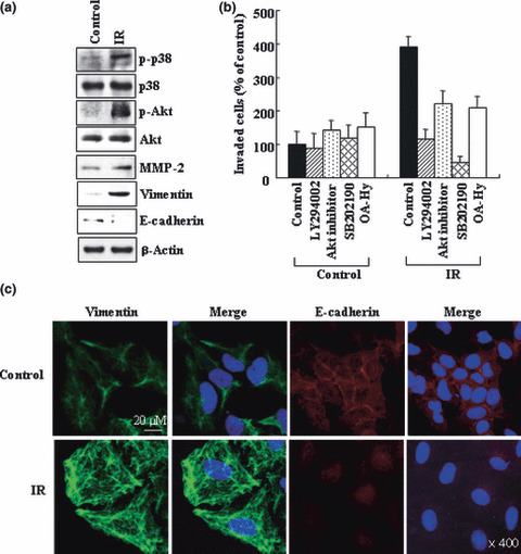
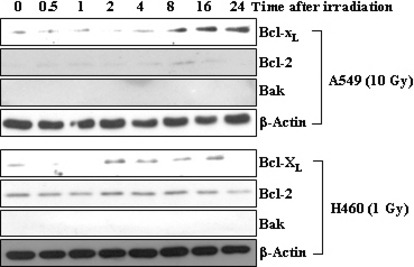
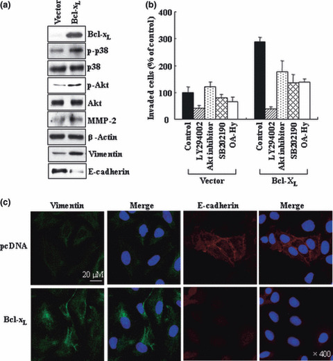
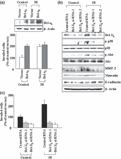
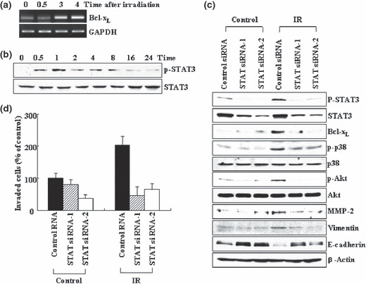
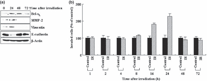
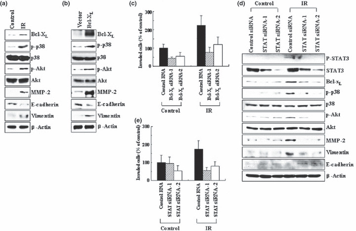
Previous reports suggest that, in addition to its therapeutic effects, ionizing radiation (IR) increases the invasiveness of surviving cancer cells. Here, we demonstrate that this activity of IR in lung cancer cells is mediated by a signaling pathway involving p38 kinase, phosphoinositide 3-kinase, Akt, and matrix metalloproteinase (MMP-2). The invasion-promoting doses of IR also increased and reduced the levels of vimentin and E-cadherin, respectively, both of which are markers for the epithelial-mesenchymal transition (EMT). Interestingly, all of these malignant actions of IR were mimicked by the overexpression of Bcl-X(L), a pro-survival member of the Bcl-2 family, in lung cancer cells. Moreover, both RNA and protein levels of Bcl-X(L) were elevated upon irradiation of the cells, and the prevention of this event using small-interfering RNAs of Bcl-X(L) reduced the ability of IR to promote invasion signals and EMT-associated events. This suggests that Bcl-X(L) functions as a signaling mediator of the malignant effects of IR. It was also demonstrated that IR enhances signal transducer and activator of transcription 3 (STAT3) phosphorylation, and the reduction of STAT3 levels via RNA interference prevented IR-induced Bcl-X(L) accumulation, and thus all the tested Bcl-X(L)-dependent events. Overall, the data suggest that IR induces Bcl-X(L) accumulation via STAT3, which then promotes cancer cell invasion and EMT-associated markers. Our findings demonstrate a novel function of Bcl-X(L) in cancer, and also advance our understanding of the malignant actions of IR significantly.


Figures

Figure 1
Figure 1
Ionizing radiation (IR) promotes malignant behaviors in A549 cells. (a) A549 cells were irradiated with 10 Gy of γ‐rays. After 24 h of incubation, cell lysates and conditioned media were prepared. Cell lysates were analyzed for p‐p38, p38, p‐Akt, Akt, vimentin, and E‐cadherin levels by western blotting using β‐actin as a loading control. The conditioned media were analyzed to compare the levels of secreted matrix metalloproteinase (MMP‐2). (b) A549 cells were incubated with or without inhibitors for phosphoinositide 3‐kinase (PI3K) (LY294002, 5 μM), Akt (Akt inhibitor I, 5 μM), p38 kinase (SB202190, 5 μM), or MMP‐2 (OA‐Hy, 5 μM) for 30 min, and then irradiated. After 24 h of incubation, cellular invasiveness was compared on Matrigel‐coated polycarbonate filters. The experiment was repeated three times, and the means and SDs were determined. (c) The control and irradiated cells were immunostained to analyze vimentin and E‐cadherin. DAPI staining to detect nuclei was also carried out. Stained cells were visualized with a confocal microscope.
Figure 2
Figure 2
Bcl‐XL is a common target of ionizing radiation (IR) in both A549 and H460 cells. A549 and H460 cells were exposed to sublethal doses of IR, 10 and 1 Gy, respectively. At the indicated times after irradiation, the levels of Bcl‐XL, Bcl‐2, and Bak were compared via western blotting.
Figure 3
Figure 3
Bcl‐XL overexpression mimics the effects of ionizing radiation (IR). (a) A549 cells were transiently transfected with empty pcDNA3 vectors or vectors containing Bcl‐XL cDNA. The levels of the indicated components were compared via western blotting. (b) The control and Bcl‐XL transfectants were incubated with or without indicated inhibitors for 30 min, then analyzed for their invasiveness. (c) The transfectants were immuno‐stained and analyzed for vimentin and E‐cadherin.
Figure 4
Figure 4
Bcl‐XL mediates the malignant actions of ionizing radiation (IR). (a) The transfectants were irradiated (10 Gy), and incubated for 24 h. Cellular levels of Bcl‐XL and cellular invasiveness were compared. (b) Control and Bcl‐XL siRNAs were introduced into A549 cells. After 24 h of incubation, the cells were irradiated, then incubated for an additional 24 h. Levels of the indicated components were compared via western blotting. (c) Cellular invasiveness was analyzed.
Figure 5
Figure 5
Signal transducer and activator of trans‐cription 3 (STAT3) mediates ionizing radiation (IR)‐induced Bcl‐XL accumulation. (a) A549 cells were irradiated. At the indicated times, the levels of Bcl‐XL mRNA were compared via RT‐PCR, using GAPDH as an internal control. (b) The irradiated cells were analyzed for levels of STAT3 protein and its phosphorylation via western blotting. (c) Control and STAT3 siRNAs were introduced into A549 cells. After 24 h of incubation, the cells were irradiated and incubated for an additional 24 h. Levels of the indicated components were compare via western blotting. (d) Cellular invasiveness was compared.
Figure 6
Figure 6
Ionizing radiation (IR) promotes invasion in a transient mode. (a) Levels of Bcl‐XL, matrix metalloproteinase‐2 (MMP‐2), vimentin, and E‐cadherin were analyzed by western blotting at the indicated times after irradiation. (b) Cellular invasiveness was compared at the times specified.
Figure 7
Figure 7
Role of Bcl‐XL and signal transducer and activator of transcription 3 (STAT3) in H460 cells. (a) H460 cells were irradiated (1 Gy), and incubated for 24 h. Levels of the indicated components were analyzed by western blotting. (b) H460 cells were transiently transfected with the control and Bcl‐XL expression vectors. Levels of the indicated components were compared. (c) Control and Bcl‐XL siRNAs were introduced into H460 cells. After 24 h of incubation, the cells were irradiated, incubated for an additional 24 h, and analyzed for invasiveness. (d) Control and STAT3 siRNAs were introduced into H460 cells. After 24 h of incubation, the cells were irradiated, incubated for an additional 24 h, and subsequently analyzed for the relevant components by western blotting. (e) Cellular invasiveness was compared.

References

    1. Schmidt‐Ullrich RK. Molecular targets in radiation oncology. Oncogene 2003; 22: 5730–3. - PubMed
    1. Barcellos‐Hoff MH, Park C, Wright EG. Radiation and the microenvironment‐tumorigenesis and therapy. Nat Rev Cancer 2005; 5: 867–75. - PubMed
    1. Kumar P, Miller AI, Polverini PJ. Ap38 MAPK mediates γ‐irradiation‐induced endothelial cell apoptosis, and vascular endothelial growth factor protects endothelial cells through the phosphoinositide 3‐kinase‐Akt‐Bcl‐2 pathway. J Biol Chem 2004; 279: 43352–60. - PubMed
    1. Park CM, Park MJ, Kwak HJ et al. Ionizing radiation enhances matrix metalloproteinase‐2 secretion and invasion of glioma cells through Src/epidermal growth factor receptor‐mediated p38/Akt and phosphatidylinositol 3‐kinase/Akt signaling pathways. Cancer Res 2006; 66: 8511–9. - PubMed
    1. Cheng JH, Chou CH, Kuo ML, Hsieh CY. Radiation‐enhanced hepatocellular carinoma cell invasion with MMP‐9 expression through PI3K/Akt/NF‐κB signal transduction pathway. Oncogene 2006; 25: 7009–18. - PubMed

Publication types