

doi: 10.1111/j.1528-1167.2009.02436.x.
MRI analysis of sulcation morphology in polymicrogyria
Affiliations
- PMID: 20331706
- PMCID: PMC3821984
- DOI: 10.1111/j.1528-1167.2009.02436.x
Item in Clipboard
MRI analysis of sulcation morphology in polymicrogyria
Epilepsia.
2010 Feb.
No abstract available
Figures

Axial T2 weighted image at a higher level shows markedly abnormal sulcation over the posterior frontal and parietal lobes, with deep infoldings (large black arrows) of dysmorphic cortex. The apparent curvilinear heterotopia in the right frontal subcortical white matter (small black arrows) was an infolding of cortex from an adjacent image.

A. Axial T1 weighted image in a patient with perisylvian polymicrogyria shows coarse cortex throughout most of both hemispheres. In addition, there are some abnormal cortical infoldings. B. Axial T1 weighted image at a higher level shows deep infoldings (black arrows) that run parallel to the overlying cortex, giving the appearance of palisades of dysmorphic cortex.

A. Axial T1 weighted image in a patient with perisylvian polymicrogyria shows coarse cortex throughout most of both hemispheres. In addition, there are some abnormal cortical infoldings. B. Axial T1 weighted image at a higher level shows deep infoldings (black arrows) that run parallel to the overlying cortex, giving the appearance of palisades of dysmorphic cortex.
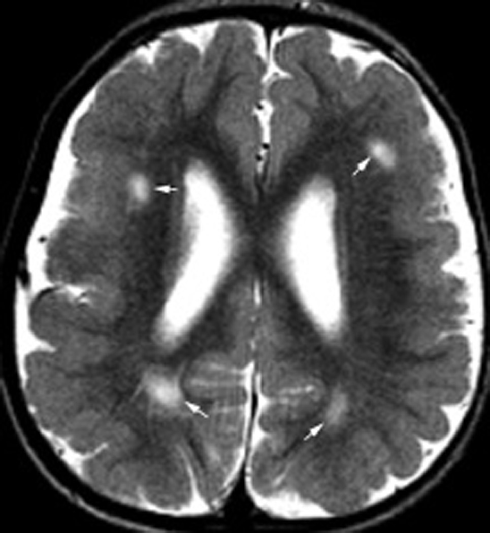
Axial T2 weighted image in an infant with developmental delay and seizures due to bilateral frontal polymicrogyria shows multiple areas (arrows) of shallow microgyri in the bilateral frontal lobes. Contrast this with the coarse appearance in Figure 1.

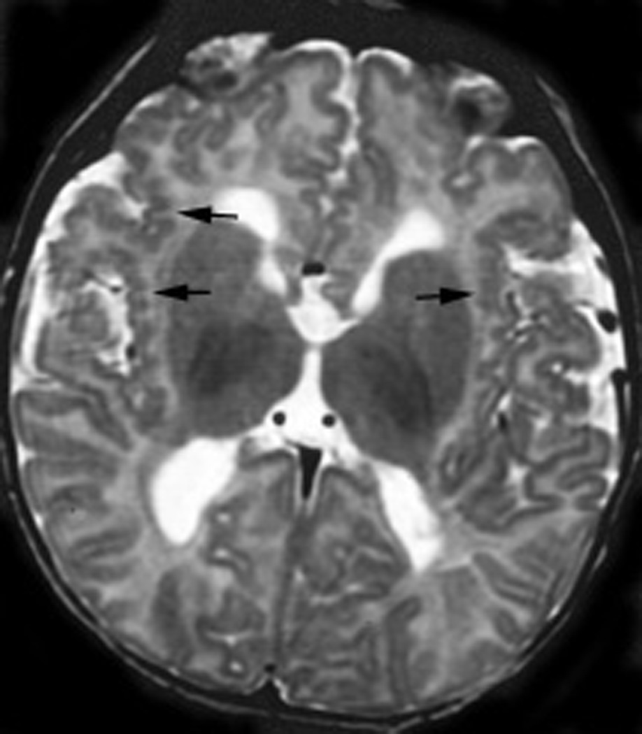
Localized delicate polymicrogyria in an infant with Aicardi syndrome. Delicate PMG with thin cortex and small, shallow sulci is present throughout much of the anterior right frontal lobe (arrows) and, to a lesser extend, left frontal lobe. The asymmetry in hemisphere size is due to ventricular cysts on the left.

Axial T2 weighted image in an infant with seizures and severe developmental delay shows multiple long, narrow gyri separated by deep sulci. On close examination, the cortex of each long, narrow gyrus is composed of many microgyri. Ventricles are enlarged and white matter is markedly diminished.

Axial T1 weighted image shows a deep infolding of thick cortex with irregular inner and outer margins, characteristic of coarse PMG.
Axial T2 weighted image shows coarse PMG that extends to the medial surfaces of the cerebral hemispheres (PMG typically relatively spares the medial and inferior surfaces of the cerebral hemispheres) and is associated with subcortical regions of abnormal myelination (also unusual for PMG).
References
-
- Bingham PM, Lynch D, Mcdonald-mcginn D, Zackai E. Polymicrogyria in chromosome 22 deletion syndrome. Neurology. 1998;51:1500–1502. - PubMed
-
- Chang BS, Apse KA, Caraballo R, Cross JH, Mclellan A, Jacobson RD, Valente KD, Barkovich AJ, Walsh CA. A famillial syndrome of unilateral polymicrogyria affecting the right hemisphere. Neurology. 2006;66:133–135. - PubMed
Publication types
MeSH terms
Grants and funding
LinkOut - more resources
Full Text Sources
Medical
