. 2010 Apr 6;107(14):6292-7.
doi: 10.1073/pnas.0913403107. Epub 2010 Mar 23.

Bluetongue virus coat protein VP2 contains sialic acid-binding domains, and VP5 resembles enveloped virus fusion proteins

Affiliations

Bluetongue virus coat protein VP2 contains sialic acid-binding domains, and VP5 resembles enveloped virus fusion proteins

Xing Zhang et al. Proc Natl Acad Sci U S A. .

Abstract

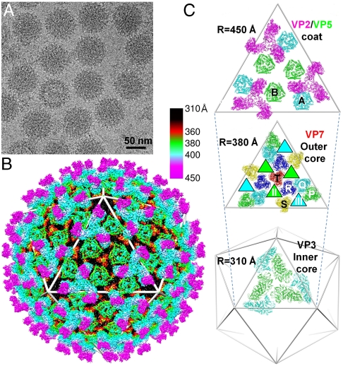
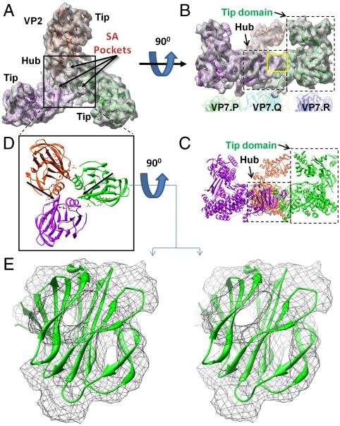
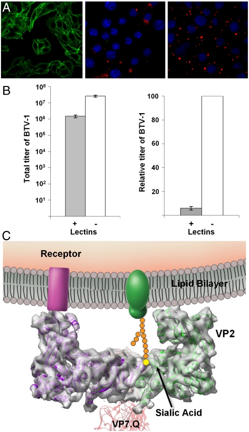
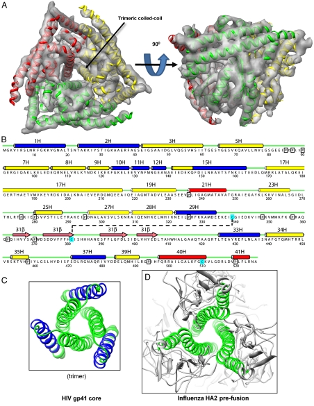
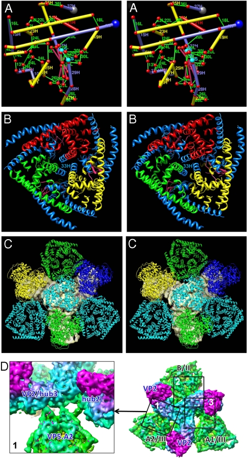
Bluetongue virus (BTV) is transmitted by blood-feeding insects (Culicoides sp.) and causes hemorrhagic diseases in livestock. BTV is a nonenveloped, double-stranded RNA (dsRNA) virus with two capsids: a well-studied, stable core enclosing the dsRNA genome and a highly unstable, poorly studied coat responsible for host cell attachment and entry. Here, based on cryo-electron microscopy (cryoEM), we report a 7-A resolution structure of the infectious BTV virion, including the coat proteins. We show that unlike other dsRNA viruses, the VP2 attachment trimer has a triskelion shape composed of three tip domains branching from a central hub domain. We identify three putative sialic acid-binding pockets in the hub and present supporting biochemical data indicating sugar moiety binding is important for BTV infection. Despite being a nonenveloped virus, the putative VP5 membrane penetration trimer, located slightly inward of the VP2 attachment trimer, has a central coiled-coil alpha-helical bundle, similar to the fusion proteins of many enveloped viruses (e.g., HIV, herpesviruses, vesicular stomatitis virus, and influenza virus). Moreover, mapping of the amino acid sequence of VP5 to the secondary structural elements identified by cryoEM locates 15 amphipathic alpha-helical regions on the external surface of each VP5 trimer. The cryoEM density map also reveals few, weak interactions between the VP5 trimer and both the outer-coat VP2 trimer and the underlying core VP7 trimer, suggesting that the surface of VP5 could unfurl like an umbrella during penetration and shedding of the coat to release the transcriptionally active core particle.


Conflict of interest statement

The authors declare no conflict of interest .

Figures

Fig. 1.
Fig. 1.
Structure of BTV-1 by cryoEM at 7-Å resolution. (A) CryoEM pictures of BTV. (B) 3D reconstruction of BTV at 7-Å resolution, color coded by radial position: outer-coat VP2 (magenta and cyan), inner-coat VP5 (green), outer core VP7 (red and black), and inner core VP3 (not visible). See also Movie S1. (C Top) Outer-coat proteins VP2 (T = 3) (magenta) and VP5 (T = 6) (B and A conformers in green and cyan, respectively). (Middle) Outer core protein VP7 (T = 13) with channels II and III (green- and cyan-filled triangles underneath B and A conformers of VP5). VP7 trimers are designated as P, Q, R, S, and T based on their locations in the asymmetric unit (8). (Bottom) Two inner core protein VP3 conformers colored cyan and green.
Fig. 2.
Fig. 2.
Fold of VP2 and identification of SA-binding pocket. Top (A) and side (B) views of the density map of a VP2 triskelion after 3-fold averaging, with three tip domains and a hub (black box) comprised of three hub domains. See also Movie S2. Each hub domain harbors a SA-binding pocket. The hub sits on a VP7.Q trimer, and each of the three VP2 tip domains sits atop its own VP7 trimer (either P, R, or S). (C) Ribbon model of (B), rich in α-helices and β-sheets. (D) Top view of atomic-resolution ribbon models of three VP8 SA-binding domains of rotavirus with positions determined by docking to the density map of VP2 of BTV. (E). Stereo view of the ribbon model of the VP8 SA-binding domain from rotavirus docked into the density map (threshold = 1.5σ) of BTV’s VP2 hub from the yellow box in (B).
Fig. 3.
Fig. 3.
Sialic acid-binding activities in BTV infection. (A) HeLa cells stained with (green) FITC-labeled WGA that binds SA (Left). Nonstructural protein 2 (red) was stained by indirect immunofluorescence, and nuclei (blue) were stained with Hoescht 33258, as described in Material and Methods, 16 h after infection with BTV in the presence (+WGA) (Center) or absence (-WGA) (Right) of excess competing WGA. (B) Total (Left) and relative (Right) viral titer 16 h after infection with (+) and without (-) competing WGA. Bars represent standard errors of four replicates (SI Text). (C) Schematic illustration of the membrane attachment of VP2 by its tip domain to a cell surface receptor and by its SA-binding domain to a cell surface glycoprotein.
Fig. 4.
Fig. 4.
Fold of VP5 and prediction of secondary structure from its amino acid sequence. (A) Sixfold averaged density map of the VP5 trimer, viewed from top and side, with an embedded ribbon model. See also Movie S3. (B) Secondary structure prediction. Predicted helices are marked by cylinders, blue (amphipathic), red (hydrophobic), and yellow (other). Predicted β-strands are marked by pink arrows, loops by thin green lines, prolines by black boxes, and cysteines by cyan boxes. The black, dashed line represents a putative disulfide bond. (C, D) Ribbon diagrams of the central coiled-coil helix bundle and a triplet of peripheral helices in fusion proteins gp41 of HIV (PDB ID code 1SZT) (24) and HA2 of influenza (PDB ID code 1HGD) (26). Blue ribbons represent amphipathic regions of α-helices; green ribbons represent nonamphipathic regions of α-helices.
Fig. 5.
Fig. 5.
Mapping of the secondary structures predicted from the amino acid sequence of VP5 to the secondary structures revealed by the cryoEM density map. (A) In this stereo, side view of a cylinder model of one monomer of VP5, α-helices are represented by thick cylinders (blue = amphipathic, red = hydrophobic, and yellow = other), β-strands by intermediate thickness cylinders (pink), loops by thin cylinders (green), and the putative disulfide bond (S-S) by a thin cylinder (cyan). (B) In this stereo, top view of a VP5 trimer, amphipathic helices are colored blue, but each of the three monomers’ nonamphipathic α-helices has its own color (red, green, and yellow). The central triplet of helices includes amphipathic helix #33H. See also Movie S4 and Fig. S3. (C) Viewed in stereo from the interior of the virus toward the outer surface of the virus, the two (cyan) VP5.A and the one (green) VP5.B conformer in the inner coat fit into the three gaps between the three legs of the solid gray VP2 triskelion in the outer coat. (The hub of the VP2 triskelion is largely obscured by the VP7.Q trimer (cyan); its three tip domains are largely obscured by the VP7.P (green), VP7.R (blue), and VP7.S (yellow) trimers.) (D) Fitting into the three gaps of the VP2 triskelion, the three VP5 penetration trimers interact with at most two hub domains. Box 1 shows three thin fingers of density connecting to two hub domains. Boxes 2 and 3 are shown in Fig. S6. See also Tables S1.

References

    1. Gibbs EP, Tabachnick WJ, Holt TJ, Stallknecht DE. U.S. concerns over bluetongue. Science. 2008;320:872. - PubMed
    1. Wilson AJ, Mellor PS. Bluetongue in Europe: Past, present and future. Philos Trans R Soc Lond B Biol Sci. 2009;364:2669–2681. - PMC - PubMed
    1. Roy P, Boyce M, Noad R. Prospects for improved bluetongue vaccines. Nat Rev Microbiol. 2009;7:120–128. - PubMed
    1. Urakawa T, Ritter DG, Roy P. Expression of largest RNA segment and synthesis of VP1 protein of bluetongue virus in insect cells by recombinant baculovirus: Association of VP1 protein with RNA polymerase activity. Nucleic Acids Res. 1989;17:7395–7401. - PMC - PubMed
    1. Stauber N, et al. Bluetongue virus VP6 protein binds ATP and exhibits an RNA-dependent ATPase function and a helicase activity that catalyze the unwinding of double-stranded RNA substrates. J Virol. 1997;71:7220–7226. - PMC - PubMed

Publication types

MeSH terms

Associated data

LinkOut - more resources