. 2010 Apr 1;70(7):2799-808.
doi: 10.1158/0008-5472.CAN-09-4136. Epub 2010 Mar 23.

Dual regulation by apurinic/apyrimidinic endonuclease-1 inhibits gastric epithelial cell apoptosis during Helicobacter pylori infection

Affiliations

Dual regulation by apurinic/apyrimidinic endonuclease-1 inhibits gastric epithelial cell apoptosis during Helicobacter pylori infection

Ranajoy Chattopadhyay et al. Cancer Res. .

Abstract

Human apurinic/apyrimidinic endonuclease-1 (APE-1), a key enzyme involved in repair of oxidative DNA base damage, is an important transcriptional coregulator. We previously reported that Helicobacter pylori infection induces apoptosis and increases APE-1 expression in human gastric epithelial cells (GEC). Although both the DNA repair activity and the acetylation-mediated transcriptional regulation of APE-1 are required to prevent cell death, the mechanisms of APE-1-mediated inhibition of infection-induced apoptosis are unclear. Here, we show that short hairpin RNA-mediated stable suppression of APE-1 results in increased apoptosis in GEC after H. pylori infection. We show that programmed cell death involves both the caspase-9-mediated mitochondrial pathway and the caspase-8-dependent extrinsic pathway by measuring different markers for both the pathways. Overexpression of wild-type APE-1 in APE-1-suppressed GEC reduced apoptosis after infection; however, overexpression of the DNA repair mutant or the nonacetylable mutant of APE-1 alone was unable to reduce apoptosis, suggesting that both DNA repair and acetylation functions of APE-1 modulate programmed cell death. We show for the first time that the DNA repair activity of APE-1 inhibits the mitochondrial pathway, whereas the acetylation function inhibits the extrinsic pathway during H. pylori infection. Thus, our findings establish that the two different functions of APE-1 differentially regulate the intrinsic and the extrinsic pathway of H. pylori-mediated GEC apoptosis. As proapoptotic and antiapoptotic mechanisms determine the development and progression of gastritis, gastric ulceration, and gastric cancer, this dual regulatory role of APE-1 represents one of the important molecular strategies by H. pylori to sustain chronic infection.


Figures

Figure 1
Figure 1
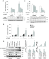
Increased apoptosis in APE-1 suppressed GEC after H. pylori infection. A, pSIREN and shRNA cells were treated with H. pylori (MOI 100) for 6, 12 or 24 h or left uninfected. Western analysis was performed for active caspase 3 (left panel) or cleaved PARP (right panel). Corresponding densitometry data normalized to α-tubulin is depicted as the mean ± SEM of three separate experiments. *, p<0.05 for shRNA cells compared to corresponding pSIREN cells. B, caspase 3 activity was measured in pSIREN and shRNA cells with or without H. pylori infection (MOI 100) for 6, 12 and 24 h. Bars represent normalized data (mean ± SEM, n=3), *, p <0.05 for shRNA cells compared to corresponding pSIREN cells. C, pSIREN and shRNA cells were treated with 10 or 20 µM of caspase 8 or caspase 9 inhibitor 1 h prior to H. pylori infection (MOI 100) for 24 h or left uninfected and analyzed by immunblotting for active caspase 3.
Figure 2
Figure 2
Reduction of APE-1 level enhances H. pylori-mediated apoptosis via the mitochondria-dependent pathway. pSIREN and shRNA cells were treated with H. pylori (MOI 100 ) for 6, 12 or 24 h or left uninfected. Western analysis was performed for (A) active caspase 9 (B) cytosolic cytochrome c or (C) Bcl-xL and Bcl-xS. For Bcl-xS and Bcl-xL normalized data are shown as a ratio. D, loss of MMP in pSIREN and shRNA cells with or without H. pylori infection (MOI 100) for 1 h. (A–D) Bars depict normalized data (mean ± SEM, n=3), *, p <0.05 for infected shRNA cells compared to corresponding pSIREN cells.
Figure 3
Figure 3
Reduction of APE-1 level enhances H. pylori-mediated apoptosis via the extrinsic pathway. pSIREN and shRNA cells were treated with H. pylori (MOI 100) for 4, 8 or 12 h or left uninfected. Western analysis was performed for (A) active caspase 8 and (B) FLIPL and FLIPS. Corresponding densitometry data, normalized to α-tubulin, are depicted as the mean ± SEM of three separate experiments. *, p<0.05 for shRNA cells compared to corresponding pSIREN cells. C, caspase 8 activity was measured in AGS, pSIREN and shRNA cells with or without H. pylori infection (MOI 100) for 4, 8 or 12 h. Bars depict normalized data (mean ± SEM, n=3), *, p <0.05 for shRNA cells compared to corresponding pSIREN cells. D, immunoprecipitation of DISC with Fas antibody at 2 and 4 h after H. pylori infection in pSIREN and shRNA cells. Western analysis was performed to detect pro-caspase 8, FADD, FLIPL and FLIPS. IgG was used as the loading control. Corresponding densitometry data normalized to IgG are depicted as the mean ± SEM of three separate experiments. *, p<0.05 for shRNA cells compared to corresponding pSIREN cells.
Figure 4
Figure 4
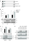
Acetylation and DNA repair functions of APE-1 both contribute to the inhibition of apoptosis. A, schematic diagram showing WT human APE-1 (upper panel), the repair mutant (middle panel) and the acetylation mutant (bottom panel). B, shRNA cell extracts were prepared after transient transfection followed by H. pylori infection and analyzed by western blot for active caspase 3. FLAG antibody was used to assess transfected APE-1 expression. Corresponding densitometry data, normalized to α-tubulin, are depicted as the mean ± SEM of three separate experiments. *, p<0.05 compared to WT APE-1. C, dose dependence of H. pylori infection in the induction of APE-1 acetylation. Densitometric analysis was performed to normalize acetylated APE-1 (AcAPE-1) to total APE-1 with an arbitrary value of 1 assigned to uninfected pSIREN cells. D, H. pylori dose-specific response of the acetylation function of APE-1 in inhibiting the intrinsic apoptotic pathway. shRNA cells were transiently transfected and infected with H. pylori (MOI 100 and 300) for 8 h (cytochrome c and Bax) and 12 h (caspase 9). Cells were lysed and analyzed by western blot for cytosolic cytochrome c, Bax and active caspase 9.
Figure 5
Figure 5
DNA repair activity of APE-1 is required to inhibit mitochondria-mediated apoptosis during H. pylori infection. A, shRNA cells were transiently transfected as in Fig. 4B and infected with H. pylori for 6 or 12 h before performing western analysis with active caspase 9 and Bcl-xL antibody. B, caspase 9 activity assay of shRNA cells identically treated to those described in A. Data are shown as fold change of each H. pylori infected group to their respective uninfected group (mean ± SEM, n=4) where the uninfected vector control is set to an arbitrary value of 1. *, p <0.05 for WT APE-1 or K6R/K7R compared to empty vector or H309N. C, measurement of loss of MMP in shRNA cells first transfected and then infected for 1 h with H. pylori (MOI 100). Bars depict fold change for each H. pylori infected group relative to uninfected controls (mean ± SEM, n=3) which is set to an arbitrary value of 1. *, p <0.05 for WT APE-1 or K6R/K7R compared to empty vector or H309N.
Figure 6
Figure 6
The acetylation function of APE-1 is required to inhibit the extrinsic pathway of apoptosis during H. pylori infection. A, shRNA cells were transiently transfected as in Fig. 4B and infected with H. pylori for 4 or 8 h before performing western analysis for active caspase 8. B, caspase 8 activity assay of shRNA cells identically treated to those described in A. Data are shown as fold change of each H. pylori infected group to their respective uninfected group (mean ± SEM, n=5) where the uninfected vector control is set to an arbitrary value of 1. *, p <0.05 for WT APE-1 or H309N compared to empty vector or K6R/K7R. C, western analysis of FLIPS in shRNA cells, transiently transfected and then infected as described previously. α-tubulin was used as the loading control. Corresponding densitometry data, normalized to α-tubulin, are depicted as the mean ± SEM of three separate experiments. *, p <0.05 for WT APE-1 or H309N compared to empty vector or K6R/K7R.

References

    1. Crowe SE. Helicobacter infection, chronic inflammation, and the development of malignancy. Curr Opin Gastroenterol. 2005;21:32–38. - PubMed
    1. Ernst PB, Peura DA, Crowe SE. The translation of Helicobacter pylori basic research to patient care. Gastroenterology. 2006;130:188–206. - PubMed
    1. Crowe SE, Alvarez L, Sherman PM, et al. Expression of interleukin-8 and CD54 by human gastric epithelium after H. pylori infection in vitro. Gastroenterology. 1995;108:65–74. - PubMed
    1. Bagchi D, Bhattacharya G, Stohs SJ. Production of reactive oxygen species by gastric cells in association with Helicobacter pylori. Free Rad Res. 1996;24:439–450. - PubMed
    1. Teshima S, Rokutan K, Nikawa T, Kishi K. Guinea pig gastric mucosal cells produce abundant superoxide anion through an NADPH oxidase-like system. Gastroenterology. 1998;115:1186–1196. - PubMed

Publication types

MeSH terms

Substances