. 2010 Apr 1;70(7):2901-10.
doi: 10.1158/0008-5472.CAN-09-3229. Epub 2010 Mar 23.

PTPH1 dephosphorylates and cooperates with p38gamma MAPK to increase ras oncogenesis through PDZ-mediated interaction

Affiliations

PTPH1 dephosphorylates and cooperates with p38gamma MAPK to increase ras oncogenesis through PDZ-mediated interaction

Song-Wang Hou et al. Cancer Res. .

Abstract

Protein phosphatases are believed to coordinate with kinases to execute biological functions, but examples of such integrated activities, however, are still missing. In this report, we have identified protein tyrosine phosphatase H1 (PTPH1) as a specific phosphatase for p38gamma mitogen-activated protein kinase (MAPK) and shown their cooperative oncogenic activity through direct binding. p38gamma, a Ras effector known to act independent of its phosphorylation, was first shown to require its unique PDZ-binding motif to increase Ras transformation. Yeast two-hybrid screening and in vitro and in vivo analyses further identified PTPH1 as a specific p38gamma phosphatase through PDZ-mediated binding. Additional experiments showed that PTPH1 itself plays a role in Ras-dependent malignant growth in vitro and/or in mice by a mechanism depending on its p38gamma-binding activity. Moreover, Ras increases both p38gamma and PTPH1 protein expression and there is a coupling of increased p38gamma and PTPH1 protein expression in primary colon cancer tissues. These results reveal a coordinative oncogenic activity of a MAPK with its specific phosphatase and suggest that PDZ-mediated p38gamma/PTPH1 complex may be a novel target for Ras-dependent malignancies.


Conflict of interest statement

Disclosure of Potential Conflicts of Interest

No potential conflicts of interest were disclosed.

Figures

Figure 1
Figure 1
p38γ requires C-terminus to increase Ras transformation and to bind PTPH1. A, p38γ requires its C-terminus to increase Ras transformation. IEC-6 cells were stably expressed with p38γ or its mutants (see left for structures) and then transduced with retrovirus containing K-Ras, which were analyzed for soft-agar growth (middle) and protein expression (right). Colony numbers from 20 fields per 60 mm dish are counted and results shown are relative over vector controls (from three separate experiments with each in triplicate, * p < 0.05 by ANOVA). B, yeast two-hybrid screening and GST pull-down reveal a PDZ-dependent interaction of p38γ with PTPH1. The detail procedure and full results of the screening were described in Supplemental data and Fig. S1A/B. The β-Gal activity assay with PTPH1 confirmed a positive reaction with p38γ (left panel) and structure of human PTPH1 protein was depicted on bottom (left). GST pull-down results were given on right panel with the input control in the middle (see Fig. S1C for similar GST and GST-PTPH1 proteins used).
Figure 2
Figure 2
The PDZ dictates p38 interaction with PTPH1 in vivo. A, a role of the PDZ motif in p38 interacting with PTPH1. Flag-tagged p38γ/α constructs were expressed with and without HA-PTPH1 in 293T cells and Flag/HA precipitates were examined by Western. Please note that Flag p38γ and Flag p38α+13 also bind endogenous PTPH1 in the absence of HA-PTPH1 (lane 2 and 3 from left). B-D, p38γ depends on its phosphorylation and C-terminus to bind the PDZ domain of PTPH1 protein. Expressed proteins in 293T cells were isolated and examined for p38γ-PTPH1 binding. Please note that Flag-p38γ fails to bind the PTPH1ΔPDZ and the bound WT PTPH1 also leads to decreased p38γ phosphorylation from the input control (B/D). Results from C showed that stably expressed Flag-p38γ (but not its Δ4 mutant) also binds endogenous PTPH1 protein in IEC-6/K-Ras cells.
Figure 3
Figure 3
PTPH1 dephosphorylates p38γ but not p38α in vitro and in vivo. A, PTPH1 dephosphorylates p38γ but not p38α in vitro. Flag-tagged p38α/γ were co-expressed with MKK6 in 293T cells and activated p38s were isolated with a Flag antibody, which were examined for in vitro phosphorylation using a specific p-p38 antibody following incubation with GST-PTPH1. The percentage indicates the relative p-p38s (normalized to Flag-p38α/γ) over those in the absence of GST-PTPH1 (measured with ImageQuant 5.0 software). B, p38γ is dephosphorylated by PTPH1 in vivo dependent of its PDZ-binding motif. Different amounts of HA-PTPH1 were co-expressed with indicated constructs in 293T cells and examined for p38γ phosphorylation. C, there is an increased complex-formation between p38γ and PTPH1/DA. Flag-p38γ was transiently co-expressed with PTPH1 or PTPH1/DA and expressed proteins were isolated in the absence or the presence of phosphatase inhibitors (1mM Sodium Vanadate, 20mMβ-glycerophosphate, and 20mM ρ-nitrophenylphosphate) and analyzed by Western. D, PTPH1 binding and dephosphorylating p38γ are both inhibited by a peptide corresponding to the p38γ C-terminus. p38γ and PTPH1 were expressed in 293T cells for 24 hr, which were then subjected to the peptide treatment (10 μM, 5 hr) and Flag IP and/or Western analyses (see Fig. S2B for input control).
Figure 4
Figure 4
PTPH1 signals downstream of Ras and p38γ and is required for Ras-dependent colon cancer growth. A, Ras increases p38γ and PTPH1 protein expression. Ras transformed IEC-6 cells were examined for protein expression (left). In addition, normal IEC-6 cells were transiently infected with LZRS retrovirus (expressing H-Ras or K-Ras) or adenovirus (p38γ) for 48 h, and examined for protein expression (middle and right). B, C, depletion of PTPH1 or p38γ protein expression inhibits soft-agar growth of HCT116 or SW480 human colon cancer cells. Cells were transiently infected with lentivirus (shLuc, #1 and #2 shRNA against PTPH1 or p38γ as indicated) and 72 h later assayed for the soft-agar growth and Western blot. The relative colony number is shown as in Fig. 1B from three separate experiments (* p < 0.05 vs. sh-Luc).
Figure 5
Figure 5
PTPH1 promotes colon cancer growth in vivo and requires its p38γ-binding activity to increase Ras-dependent growth in vitro. A, PTPH1 depletion inhibits the tumor growth in mice. HCT116 cells were stably infected with Lenti-shLuc or Lenti-#1shPTPH1 in cell culture (see Fig. S4A for Western) and 2 × 10⁶ of these cells s.c. injected into both front flanks of nude mouse (right, shLuc; left, shPTPH1) and the tumor growth was monitored. Moreover, tumors were excised, photographed, and weighed at the end of experiment (insert, p < 0.05 between two groups for either tumor weight or volume in all time point) and similar results were obtained from additional two experiments (Fig. S3A/B). B, C, PTPH1 requires its PDZ domain to increase Ras transformation. IEC-6/K-Ras cells were stably expressed with PTPH1 or its PDZ-deleted mutant PTPH1ΔPDZ, and subjected to Western (B) and Soft-agar assays (C) (means of three separate experiments, * p < 0.05). D, disruption of the p38γ-PTPH1 interaction inhibits colon cancer cell proliferation. HCT116 cells were incubated with the wild-type and mutant peptide as described in Fig. 3D with a TAT-GFP as a separate control and cell growth was estimated by thymidine incorporation as previously described (10). Results shown (relative to TAT-GFP control) are mean of three separate experiments (*, p < 0.05, p38γC13 versus p38γC13A).
Figure 6
Figure 6
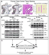
Roles of PTPH1 and p38γ in colon cancer. A, hyperexpressed p38γ correlates with increased PTPH1 expression in primary colon cancer tissues. p38γ and PTPH1 protein expression in invasive colon carcinomas were analyzed by immuno-histochemistry staining. Representative pictures from the same patient were given in left two panels showing an increased positive brown signals for p38γ and PTPH1 in malignant tissues (indicated by an open arrow head) over those in normal gland (marked with a closed arrow head). A Pearson’s correlation was reached between increased p38γ and PTPH1 protein expression in this group of specimens (142 cases) after subtracting signals of the tumors from those of matched normal tissues (p < 0.05, right panel). Additional results about p38γ/PTPH1 protein expression in colon cancer specimens are shown in Fig. S4C and Table S1. B, levels of endogenous PTPH1 protein expression inversely couple with intrinsic p38γ phosphorylation. Cells were depleted of PTPH1 protein and subjected to IP/Western analysis for increased p-p38γ proteins (IP) (left). On right panel, protein samples were prepared from two pairs of tumors (#1 and #4 mouse, Fig. 5A insert) and subjected to the IP/WB analysis, which together with another set of experiments (Fig. S3C) showed a coupling of decreased PTPH1 protein expression with increased p38γ phosphorylation. * indicates a fold increase in p-p38γ over individual shLuc control. C, an experimental model shows a PDZ-mediated cooperative oncogenic activity of p38γ MAPK with its phosphatase PTPH1. Ras was shown to increase protein expression of p38γ and PTPH1 in which PTPH1 is also induced by its substrate p38γ and acts to dephosphorylate p38γ through PDZ-mediated binding. Experimental evidence is presented to indicate that it is the PDZ-mediated p38γ/PTPH1 complex that possesses cooperative oncogenic activity leading to increased colon cancer growth. The dotted lines indicate that p38γ and PTPH1 may be up-regulated by Ras-independent proliferative signals.

Similar articles

Cited by

References

    1. Downward J. Targeting Ras signalling pathways in cancer therapy. Nature Rev Cancer. 2003;3:11–22. - PubMed
    1. Han J, Sun P. The pathways to tumor supression via route p38. Trends Biochem Sci. 2007;32:364–71. - PubMed
    1. Loesch M, Chen G. The p38 MAPK stress pathway as a tumor suppressor or more? Front Biosci. 2008;13:3581–93. - PMC - PubMed
    1. Chen G, Hitomi M, Han J, Stacey DW. The p38 pathway provides negative feedback to Ras proliferative signaling. J Biol Chem. 2000;275:38973–80. - PubMed
    1. Qi X, Tang J, Pramanik R, et al. p38 MAPK activation selectively induces cell death in K-ras mutated human colon cancer cells through regulation of vitamin D receptor. J Biol Chem. 2004;279:22138–44. - PubMed

Publication types

MeSH terms

Substances