Review
. 2010 Mar 23;3(114):pe10.
doi: 10.1126/scisignal.3114pe10.

ER calcium and Alzheimer's disease: in a state of flux

Affiliations
Review

ER calcium and Alzheimer's disease: in a state of flux

Mark P Mattson. Sci Signal. .

Abstract

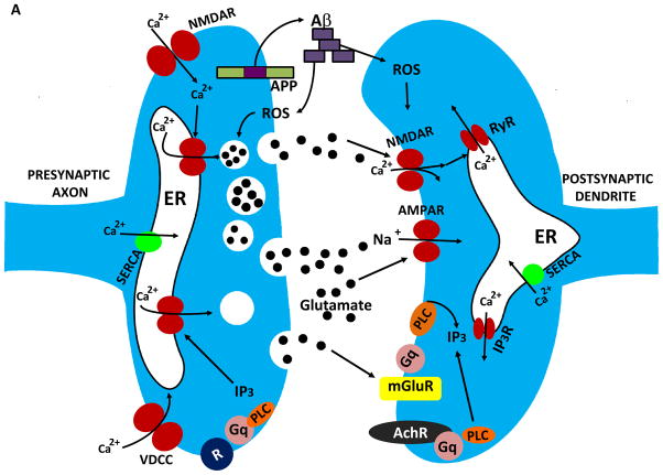
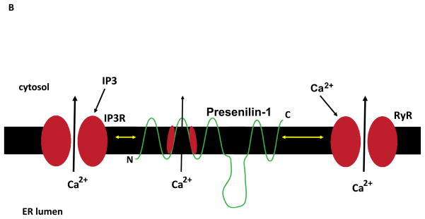
The calcium ion (Ca(2+)) plays fundamental roles in orchestrating dynamic changes in the function and structure of nerve cell circuits in the brain. The endoplasmic reticulum (ER), an organelle that actively removes Ca(2+) from the cytoplasm, can release stored Ca(2+) through ER membrane receptor channels responsive either to the lipid messenger inositol trisphosphate (IP(3)) or to cytosolic Ca(2+). Emerging findings suggest that perturbed ER Ca(2+) homeostasis contributes to the dysfunction and degeneration of neurons that occurs in Alzheimer's disease (AD). Presenilin-1 (PS1) is an integral membrane protein in the ER; mutations in PS1 that cause early-onset inherited AD increase the pool of ER Ca(2+) available for release and also enhance Ca(2+) release through ER IP(3)- and ryanodine-sensitive channels. By enhancing Ca(2+) flux across the ER membrane, PS1 mutations may exaggerate Ca(2+) signaling in synaptic terminals and thereby render them vulnerable to dysfunction and degeneration in the settings of aging and amyloid accumulation in AD.


Figures

Figure 1
Figure 1
Mechanisms that regulate synaptic calcium dynamics, and their possible roles in the pathogenesis of Alzheimer’s disease (AD). A. The endoplasmic reticulum (ER) is present in both presynaptic axon terminals (left) and postsynaptic dendrites (right) where it regulates Ca2+-mediated processes involved in synaptic transmission and structural plasticity. In response to membrane depolarization, Ca2+ enters presynaptic terminals through voltage-dependent Ca2+ channels (VDCC) and N-methyl-D-aspartate (NMDA) glutamate receptors. The elevated cytosolic Ca2+ then triggers Ca2+ release through ryanodine receptors (RyR), and Ca2+ may also be released through IP3 receptors (IP3R) in response to activation of metabotropic receptors (R). The elevated Ca2+ concentration in the cytosol triggers fusion of glutamate-containing synaptic vesicles with the synaptic plasma membrane resulting in release of glutamate into the synaptic cleft. The β-amyloid precursor protein (APP) is present in the presynaptic membrane, where it can be proteolytically processed to release the amyloid β-peptide (Aβ). Aβ then self-aggregates on pre- and post-synaptic membranes where it causes oxidative stress and membrane lipid peroxidation which can disrupt cellular Ca2+ homeostasis. Glutamate binds postsynaptic AMPA receptors resulting in sodium influx, membrane depolarization and calcium influx through NMDA receptors and VDCC. Glutamate also binds metabotropic receptors (mGluR) coupled to the GTP-binding protein Gq which then activates phospholipase C (PLC) resulting in generation of IP3. SERCA, sarco- (smooth-) endoplasmic reticulum Ca2+-ATPase. AMPAR (alpha-amino-3-hydroxy-5-methyl-4-isoxazolepropionic acid receptor). B. Presenilin-1 (PS1) is an integral membrane protein that may modulate ER Ca2+ dynamics, thereby playing roles in synaptic plasticity and the pathogenesis of AD. PS1 can interact with IP3R and RyR, and mutations in PS1 that cause early-onset inherited AD enhance the amount of Ca2+ released in response to IP3 or Ca2+ influx. PS1 may itself function as a Ca2+ leak channel and PS1 mutations may compromise this function of PS1 resulting in an increased intraluminal Ca2+ concentration.
Figure 1
Figure 1
Mechanisms that regulate synaptic calcium dynamics, and their possible roles in the pathogenesis of Alzheimer’s disease (AD). A. The endoplasmic reticulum (ER) is present in both presynaptic axon terminals (left) and postsynaptic dendrites (right) where it regulates Ca2+-mediated processes involved in synaptic transmission and structural plasticity. In response to membrane depolarization, Ca2+ enters presynaptic terminals through voltage-dependent Ca2+ channels (VDCC) and N-methyl-D-aspartate (NMDA) glutamate receptors. The elevated cytosolic Ca2+ then triggers Ca2+ release through ryanodine receptors (RyR), and Ca2+ may also be released through IP3 receptors (IP3R) in response to activation of metabotropic receptors (R). The elevated Ca2+ concentration in the cytosol triggers fusion of glutamate-containing synaptic vesicles with the synaptic plasma membrane resulting in release of glutamate into the synaptic cleft. The β-amyloid precursor protein (APP) is present in the presynaptic membrane, where it can be proteolytically processed to release the amyloid β-peptide (Aβ). Aβ then self-aggregates on pre- and post-synaptic membranes where it causes oxidative stress and membrane lipid peroxidation which can disrupt cellular Ca2+ homeostasis. Glutamate binds postsynaptic AMPA receptors resulting in sodium influx, membrane depolarization and calcium influx through NMDA receptors and VDCC. Glutamate also binds metabotropic receptors (mGluR) coupled to the GTP-binding protein Gq which then activates phospholipase C (PLC) resulting in generation of IP3. SERCA, sarco- (smooth-) endoplasmic reticulum Ca2+-ATPase. AMPAR (alpha-amino-3-hydroxy-5-methyl-4-isoxazolepropionic acid receptor). B. Presenilin-1 (PS1) is an integral membrane protein that may modulate ER Ca2+ dynamics, thereby playing roles in synaptic plasticity and the pathogenesis of AD. PS1 can interact with IP3R and RyR, and mutations in PS1 that cause early-onset inherited AD enhance the amount of Ca2+ released in response to IP3 or Ca2+ influx. PS1 may itself function as a Ca2+ leak channel and PS1 mutations may compromise this function of PS1 resulting in an increased intraluminal Ca2+ concentration.

Similar articles

Cited by

References

    1. Alzheimer’s Association. Alzheimer’s disease facts and figures. Alzheimers Dement. 2009;5:234–270. - PubMed
    1. Fjell AM, Walhovd KB, Fennema-Notestine C, McEvoy LK, Hagler DJ, Holland D, Brewer JB, Dale AM. One-year brain atrophy evident in healthy aging. J Neurosci. 2009;29:15223–15231. - PMC - PubMed
    1. Arumugam TV, Gleichmann M, Tang SC, Mattson MP. Hormesis/preconditioning mechanisms, the nervous system and aging. Ageing Res Rev. 2006;5:165–178. - PubMed
    1. Cohen AD, Price J, Weissfeld L, James J, Rosario B, Bi W, Nebes R, Saxton J, Snitz BE, Aizenstein HA, Wolk DA, Dekosky ST, Mathis CA, Klunk WE. Basal cerebral metabolism may modulate the cognitive effects of Abeta in mild cognitive impairment: an example of brain reserve. J Neurosci. 2009;29:14770–14778. - PMC - PubMed
    1. Mattson MP. Pathways towards and away from Alzheimer’s disease. Nature. 2004;430:631–639. - PMC - PubMed